Tuesday, January 23, 2018
I am who I say I am
I am who I say I am. According to the earliest passport I have, my name then was Louisa Churchill Orrock, born on the 7th August 1958. My mother's maiden name was Hiers, and I changed my middle name by deed poll in 2012 to Hiers, although I may change it back. I say this in case my name appears on no official registers. I am a former FE A level Government and Politics lecturer but started writing about disease and the microscope after guessing about disease in 2012 and after applying for and obtaining voluntary redundancy at work. Although a US passport holder, I currently live mainly in the UK, in London.
Sunday, January 21, 2018
"Only looking at lens and biscuit wrappers under the microscope!", someone might say, which is mainly true since I am trying to establish magnification and complexity, but I will look at other things, including the butterfly wing that came with the Prinz, to establish that what one sees doesn't resemble the structure of the cell, if it would otherwise be visible at less than 800x (which is perhaps why most cells are not).
No, 40x is the strongest objective - so 800 stated magnification is correct. I was remembering that initially I thought I could see 600 x with those lenses, later more like 450, although accuracy is difficult because: (i) you can't use a slide - the lens won't go down far enough so you have to sellotape something to the microscope stage - which makes manipulation of the image difficult; (ii) manipulation of the image, pulling it through several screens from one edge to the other of the strip of wrapper became more difficult over the month I was observing because the image became more luminous at 800 and also part of the screen blurred, so that it became more difficult to find 'landmarks' in order to measure; (iii) you are also estimating the width of the screen within the cylinder, since you are attempting to establish the veracity of the magnification of the width of the lenses and what you are looking at (a screen within the cylinder? a magnified objective lens tube); even comparing the screen one sees repeatedly with circles of various sizes next to the microscope it is difficult to get a consistent estimation - ie, does the screen look to be 110 mm wide or 90 mm and is this consistent with the apparent magnification of objects within the screen when one compares them with the object itself, or would that suggest a larger or smaller screen?
Does the microscope achieve high enough magnification to view the ‘cell’?
“So the physicists have produced a microscope known as the Electron Microscope which does not use light rays at all but operates with cathode rays. Instead of a beam of light it employs a stream of electrons, and electromagnets instead of lenses.” Kenneth M. Smith, Beyond the Microscope, 1945.
“[I]n 1904, Arthur Balfour announced on the part of British science that the human race without exception had lived and died in a world of illusion until the last year of the century.” The Massachusetts Historical Society, The Education of Henry Adams, An Autobiography, September 1918.
Introduction
Although cancer was apparently diagnosed before the invention of the electron microscope, diagnosis since at least the 1960s has relied upon it. I have had no access to an electron microscope but was initially sceptical that the traditional light and lens microscope viewed what was beyond the lens or, if it did, that it could achieve anything like the higher magnifications claimed. I then saw that high magnification was apparently possible with a traditional microscope, as much as 600x with the Apex Learner. However, because of distortions at low magnification with magnifying lenses I thought that the light microscope must then be an electron microscope, neither microscope in the sense of replacing light with beams of electrons, but because they must allow remote accessing and the construction of an image unless, in the case of the light microscope, a kaleidoscopic effect is produced mechanically within the microscope. However, I have not yet ruled out the possibility that I am seeing a significantly enlarged image of the object itself rather than a reconstruction and that the complexity and focus is the result of better illumination, as is implied by the name ‘light’ microscope.
However, the fact that the magnifications are not those claimed by the manufacturer, that some of the complexity and magnification appears, in fact, to be light distortion and that I have not yet obtained consistency in terms of the size of the object as a whole and its individual parts, that we are unable to measure enlargement precisely because we are estimating the size of the screen and the extent to which the object exceeds it, that the increase in complexity at high magnification seems less suggestive of underlying structure if we examine the object with the eye alone under strong light, that the measurement of magnification for diagnostic purposes is in length only so that the highest magnification the light microscope is capable of is nevertheless insufficient to diagnose disease, the implausibility of electron beams and other mistakes in explanations of magnification, including the fact that we are not viewing the object directly with lens magnification but, from my observations, are seeing a reflected image, has encouraged me to think I am right that microscopes do not view cells, even if the conclusion may be less obvious than if it could be demonstrated that the light microscope were not viewing the object on the slide or enlarging much more than can two magnifying lenses.
The following is based on observations using the Prinz 2801 (a standard two-lens light microscope), the Apex Learner (some modification to the standard shape), and the Sunagor MagnaScope (a two-part microscope consisting of a zoom and magnifier with a funnel to direct light). Each of these are light microscopes that depend on battery or mains, mirror or natural lighting and magnifying lenses to achieve enlargement. Until recently I had underestimated what could be seen using the light microscope because I had depended only upon the Prinz 2801 and Sunagor MagnaScope, both of which relied on natural lighting after damage, one of which could apparently not see beyond the microscope, even if it showed quite complicated and ‘cell’ like structures on the screen, and the other which could, but with magnification of what looks like only around 15 times in length, but with only natural lighting, the light unit having snapped off soon after I acquired it.
However, the Apex Learner, which is illuminated either from above or below the slide, achieves surprisingly high magnifications. With the Apex Learner I appear to see a two-millimetre wide piece of transparent bronze film – the ocular lens wrappings - magnified so far by as much as 600 times –360,000 times if we think of magnification instead in terms of area, although pathologists do not. However, it is impossible to assess the actual magnification since we are judging the width of the screen we see within the cylinder rather than an image projected onto a screen (and for this reason too it can be difficult to obtain consistent results). However, magnification appears to reveal structures that cannot be seen by the eye alone, although with the object observed (the bronze-coloured wrapper) these appear to be a pattern of opaque and translucent (reversed if we light from below rather than above) rather than an underlying structure.
When I obtained the higher magnifications I did not doubt that microscopes did not diagnose disease, which I have been certain of since guessing in late 2012, but was not sure how to account for it. The telescope contains mirrors, but whether or not there are mirrors inside the microscope – and I have so far been unable to take any of them apart - these would not, from observation of what one sees , for example, if one places a mirror between two magnifying glasses, add significantly to enlargement Instead, I wondered first why it would have been necessary to have invented the implausible-sounding electron microscope rather than continue to use the light microscope for diagnosis of disease unless it were to discourage amateurs and then whether what was being implied by the name was not in fact beams of electrons providing illumination but that all microscopes are electron microscopes in the sense that they resemble other electronic communications devices: the outline of the image or the object or stain itself is detected, enlarged and filled with a constructed complex structure.
The microscope as a diagnostic instrument
The sections following observations from the Apex Learner are sceptical with respect to traditional accounts of magnification but have been influenced by hints in these as well as in other non-fiction and literature. There appear to be mistakes in standard accounts of magnification. For instance, we are viewing a reflected image rather than the object itself, so that what is actually happening within the lens is re-reflection, determined by the character of the lens and illumination. The highest magnifications I have obtained are also lower than those claimed by the manufacturer, even if surprisingly high. The highest magnification possible with the light microscope, according to the department of biological sciences at Leeds University is 1400 times, less than that needed for cell pathology. However, given that this is almost 2 million times if one thinks of enlargement in terms of area, one wonders if the definition of magnification in terms of length was not intended to prevent self-analysis of cells.
Accounts of the electron microscope, on the other hand, assert that higher magnification and resolution, or clarity, can be obtained with electron beams and magnets, with magnifications of as much as 7 million times. [encyclopedia of biology] The mechanisms chosen, electron beams and magnetic coils, may be intended to undermine criticism of diagnosis, as might also the subject of light waves, or to encourage it, although the name may also be chosen to imply that what we understand to be a new kind of microscope may instead be remote accessing and construction of the image.
Dorland’s Medical Dictionary defines magnification as “apparent magnification under the microscope”. We can see very small objects – such as mould spores – with the eye alone, while the eye itself can achieve relatively high magnification: the structure of the cornea of the eye is revealed when we squint into light. Also, with limited magnification, especially but not only when using a zoom lens, we can have the impression of significant magnification when, if we actually compare the size of what we are viewing with the size of the object itself, we realise there is little or no actual increase in size: the illusion is created by viewing an object that is further away. Also, with a magnifying glass we may notice that it is the eye itself that is doing the magnification: for example, if we place a lens directly above an object on a page and then hold the lens closer to our eyes, the object will appear larger than when viewed without the lens but so may the lens itself, ie, the object may occupy the same proportion of the lens, although usually it will occupy somewhat more . Nevertheless, the light microscope does also appear to achieve surprisingly high magnification when we compare it to a camera that blurs at limited magnification (from observation, as little as 2 to 5 times, depending upon what we are viewing and certainly more than is possible with the eye alone. Dorland’s phrase may be referring to the fact that it is impossible for the microscopist to measure magnification in the traditional microscope - without taking it apart, which I have not been able to do with the Prinz or the Apex Learner - because we do not have certainty as to the width of the screen we are viewing, whatever the stated or calculated (ie, taking into account convexity, refraction, positions and size of lenses) magnification of the ocular lenses and the width of the objective lens tubes. Or he may be referring to the character of the electron microscope. Although I have as yet no knowledge of electronics, whether or not the magnetic coil detects the object or its reflection, while any subsequent increase in size is enlargement whether or not it is a reconstruction, the complexity is added, presumably as a result of external accessing and manipulation rather than from inside the microscope, so that we are therefore not viewing an enlarged image of the object on the screen. This would be consistent with the fact that we appear only to achieve relatively limited clear enlargement with light projection and magnifying lenses.
All enlargement using light projection or lenses results in loss of clarity. This is self evident: although we can bring an object into focus that is otherwise too far away or too close to bring clearly, any enlargement will weaken in the sense of blurring the image. With light projection, we may be able to view a slide significantly beyond its size but the image will be less clear than the object at the same size and blurring and loss of visibility occur as the size increases or because of insufficient light at greater distances. The image is dissipated, or becomes less clear, with projection because, as with any enlargement, the space between outlines increases but is also distorted because the angle at which the object is projected is smaller nearer the centre. It disappears when the lines that define objects have disappeared or when the light can no longer reach far enough or is too diffuse. When using a camera we can bring an object closer but often not enlarge without loss of clarity or, much beyond 2 – 5 x (depending on what we are viewing), identify objects. A zoom may allow us to see an object in the distance relatively clearly. However, even without enlargement beyond the actual size, we will see a less clear image than the original because the effect of a glass lens is to reduce rather than increase clarity, which we can observe if we were to move closer to the object and view it at normal focal length, or if we compare the image obtained with a zoom with that obtained if we improve focus by cupping our hands in front of our eyes.
“1. (in optics) A defect in the image formed by a lens or curved mirror.” Entry for ‘abberration’ in Oxford Dictionary of Science, page 1.
Whereas with slide projection, although there is an increasing loss of clarity, the image we see increases in size as we move the object from the slide, with a magnifying lens (or microscope) we are not attempting to project an image beyond its location and there is not an indefinite advantage in placing the object at an increasing distance from the lens although magnification also increases as we remove our eye from the lens as though projection of the object is continuing beyond the lens, although as when we think the objects in a mirror change when we look at it from a different angle but they do not, so the reflected image remains the same whatever our perception of it. This needs further consideration but can in part be explained by the fact that although the lens appears to remain the same size the size of the screen with a larger number of objects on it appears much larger when our eye is against the eyepiece, although not entirely since the actual size of the individual object on the screen appears greater when viewed from a distance. It is as though the convexity of the eye itself were resulting in a smaller area being reflected onto the eye, the eye itself mimicking the action of the lens and compounding magnification, but this will need further consideration.
With lens magnification, we get blurring, loss of visibility and distortions with enlargement, and the object then becomes smaller as the effect of convexity reverses and recedes. With convex lenses, the distorting effect is also greater than with enlargement from projection alone because the angle of enlargement is determined by curvature as well as illumination. We can see the distorting effect of lenses if we look at ourselves in a slightly curved mirror. Reflection - which the next section explains is what is actually taking place with lens magnification - is usually onto a three dimensional object (the lens) whose surface is different from that of the object being viewed, unless it is a similarly shaped sphere, the distorting effect being greater with greater magnification. The three dimensional nature of the lens also increases relative flattening at the edges. Other distortions include: uneven magnification and distortion of shape, light distortion, the image turning upside down, objects being displaced from left to right or vice versa, multiple images, increasing opacity, a tunnelling effect, and will be considered after discussion of what is occurring when lens magnification takes place, after a summary of observations from the Apex Learner.
“If well corrected lenses are used, the magnifying power of the microscope should be at least that necessary to reveal the finest details resolvable by the objective. For the normal eye, this is equivalent to about 500 to 700 times the numerical aperture of the objective.” [1931]
Initial observations with the Apex Learner can be viewed in the Appendix but what follows are more recent observations where I obtained clear images approaching the magnifications claimed. My aim has been to establish the extent of magnification, although I intend once I have obtained consistent results to turn to the character of the image. Because one is estimating the size of the screen and objects on it and because of the difficulty of manipulating the image to then measure with accuracy the size of the object (a piece of bronze-coloured translucent film – the wrapping for the lenses - cut first to approximately 1 mm in width and then to 2 mm and placed under a slide casing), I have repeated the observations in an attempt to obtain consistency and to avoid the temptation to underestimate magnification and record true results.
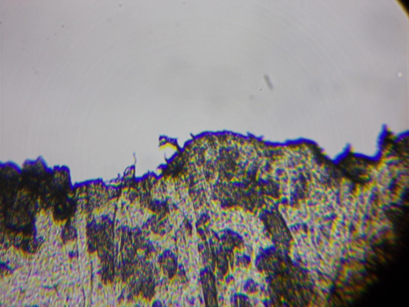
(i) First, carrying out the observation with a piece of bronze translucent film placed horizontally on the slide, and cut fairly consistently to about 2 mm in width, the size of the width of the foil on a screen appearing to be about 100 mm appeared to be 50 mm, 120 mm and 450 for the three objective lens tubes with a 10x eyepiece lens, supporting magnification of 25x, 60x, and 225x. Manipulation was difficult because the image appeared less differentiated although no less complex than with a previous piece of film (see Appendix 1). Nevertheless, we see within the ribbon thousands of tiny parts, if not completely clearly, resembling something like the map of a city. Of interest in this observation was that I noticed that with illumination from below one saw the image in negative: which is consistent with the structure reflecting patterns of light and opaque. Magnification of 25x, 60x and 225x differed from the stated magnification of 40x, 100x and 400x, and further observations will be undertaken to establish whether the variance is real.
(ii) Carrying out the observation with the same piece of film - 2mm and the 20x eyepiece, the width of the screen appeared to be about 150 mm rather than 200 mm, but it was difficult to make the judgement. With the 4x objective lens tube , the width of the ribbon appeared to be at least 150, which would indicate a magnification of 75x rather than 80 x, an insignificant variance given the difficulty of estimating the width of the screen. With the 10 x objective lens, the width of the piece of film appeared to be about 300 mm, which would indicate a magnification of 150x. With the 40x objective lens, it was not possible to position the lens tube so that the film could be viewed in focus. Instead, I cut another piece of film and placed that on the stage without a slide or casing. Then the width of the magnified object did appear to be about 4x that of the same object (ie, a part of the film, given the difficulty of manipulating and keeping in focus the ribbon when not in a casing – and whether or not the apparently shifting image was or not the result of any sort of external manipulation, this was when I first thought that ‘electron’ microscope might be code for remote viewing of all microscopes). This would indicate a magnification of at least 600x (rather than 800 times). At this highest magnification we see a similar and remarkably complex image with the thousands or tens or hundreds of thousands (repeated observation may establish which) small parts, mostly roundish, some more rectangular, as well as wider lines, larger than at lower magnifications but not obviously otherwise clearer or more complex (apart from the more rectangular shapes being more visible than at lower magnifications) or more numerous. The variance between the magnification obtained with the Apex Learner and what I had seen with magnifying glasses seemed hard to account for. The image is also remarkably still at high magnification. This has led me to believe that although the light and lens are able to detect the shape of the object, the enlargement of the outline is a reconstructed one and the complexity added, so that the Apex Learner must be an ‘electron’ microscope.
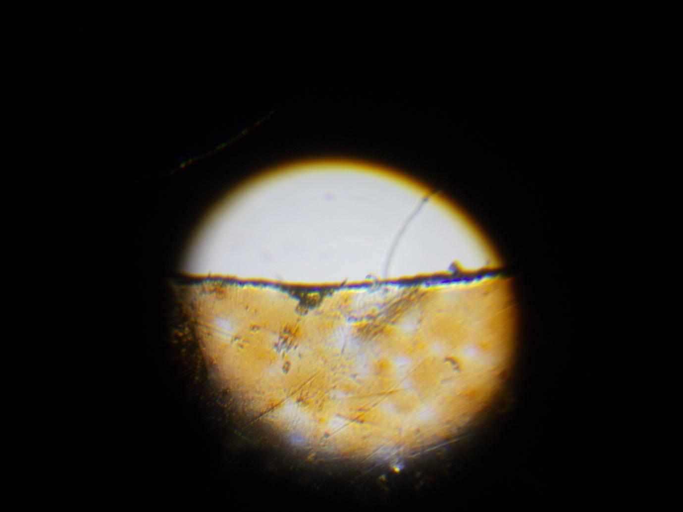
(iii) Second, carrying out the observation with a piece of Jaffa cake wrapping 2 mm wide [added subsequently from memory so that I have changed the estimation of magnification that follows; I lost the two bits of film I was working with but think the biscuit wrapping was not 4 mm, which the initial estimation suggested], I found magnification of approximately 2-400x in length or 4-16,000 times in area, which is said to be the magnification needed to view the HIV cell; this was achieved with the 10x ocular lens and 40x objective lens and so may have reached the 400 x claimed. I estimated the screen width to be about 80 mm and the piece of foil to be about 5 times the width of the screen. During this observation I was less convinced than in the previous observation that there was either remote manipulation or even a kaleidoscopic internal adjustment of the image, even though the image was not entirely clear as I moved it. Nor did the stillness of the image seem as suspicious as when I had carried out the observations with the lens wrapper. This then seemed a remarkably clear image given the magnification, even if the increase in magnification of individual structures seemed more apparent than real in terms of the actual increase in length as opposed to area. At 4x I saw the foil cover most of the screen so that the stated increase from 40x to 400x was in fact by about five rather than one hundred times. If I hold the wrapper up to the light I can just see the edge of the wrapping - where I cut it. I can also see cracks in the wrapping and what look like dust particles that may account for some of the complexity. Comparing this with what one sees with a magnifying glass of 1.5 or 2. I can start to see, especially with strong illumination, that the complex structure is discernible, the lens wrapper particularly looking something like a layer of skin. While on the one hand it does not look like, for instance, the creases one can see with the eye alone have been magnified, say, 400 times, I cannot yet say with certainty that I am overestimating the size of the screen or that the complexity is constructed or reproduced, particularly where the width of the foil covers less than the screen. What I can say is that the magnifications claimed are fairly inaccurate, whether length or area, and that this seems odd, given that it does seem to achieve high magnification. and that the resemblance of the image under the microscope to what one sees when holding the film up to strong light may support significant enlargement but not the revelation of an underlying structure.
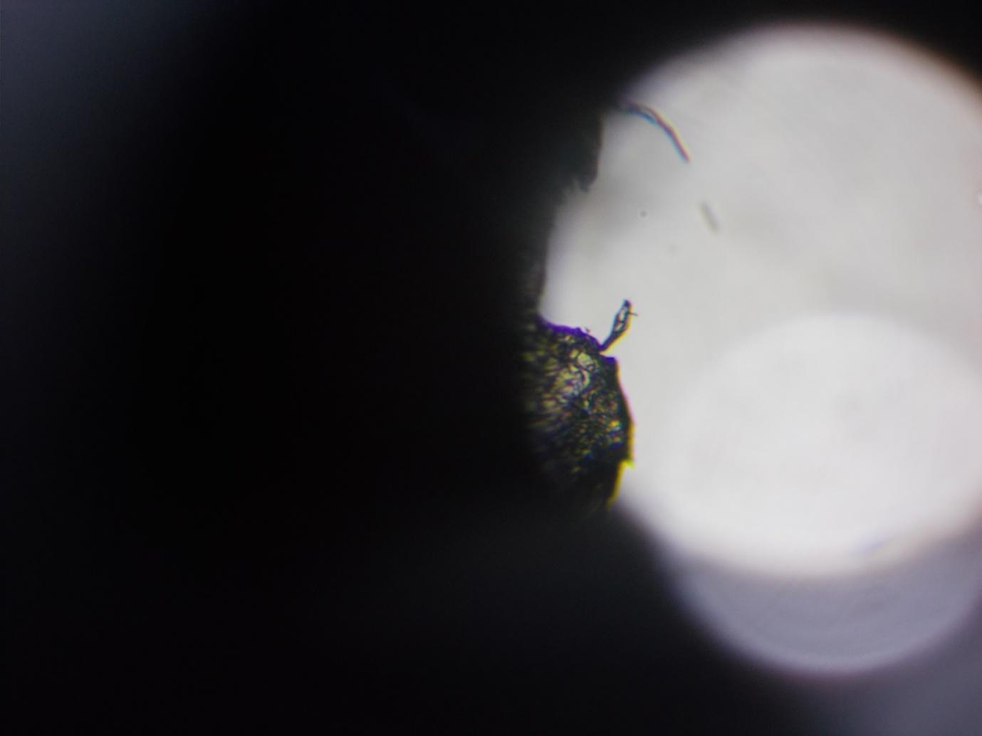
(iv) Repeating the observation again with the biscuit wrapper and the 20x eyepiece lens, what looked like a 2 mm long crack in the piece of wrapper was magnified to what looked like 40 mm with the 20 x lens and 4x objective (20x) but assuming a somewhat smaller - 60-80 rather than 80-100 than with the 10x lens - rather than larger screen than with the lower strength ocular lens. With the 10 x objective, the same crack was visible bus appeared to take up more than the screen, about 100. The length of the crack appeared to increase around 1.5 times, implying a magnification of between 30x (assuming the lower magnification was correct) and 50x. This is less than the 80 and 200x claimed by the manufacturer, although a lot more if the magnification were based on area, which it seems to be with the Sunagor MagnaScope. Although the stillness of the image is compatible with our seeing the object rather than a construction, neither on the other hand at clear magnification of 50x or 2500 x are we seeing any moving parts. With the 40x lens, interestingly, one can see that the widened edge of the wrapping is, in fact, distortion that, depending on the position of the lens, can be present or absent without loss of clarity of the rest of the image. At this magnification as with the 10x and 40x, what I take to be imperfections and particles on the eyepiece lens, are visible. I am less certain than I am with magnifying lenses that I am seeing a reflected image. However, whether or not the apparent difference between the glasses and the microscope is better lighting, notably in the case of the Apex Learner, from the round shaped light below the microscope stage, although we see more clearly and perhaps in more detail with the stronger of the two lights, the size of the outline is the same whichever of the two lights is used and also with natural lighting, where, however the detail is more difficult to see, which does not support the idea that we are seeing the underlying structure of the cell with the microscope. It also seems unlikely that however bright and guided the lighting that we would be able to see through the cylinder beyond the lenses rather than that the image is somehow captured in the lenses themselves. If it is thought unlikely that we would be able to see so clearly within a lens, the magnified structures or imperfections in the eyepiece lens are clear when the light is on and there is no object present, as is the image we see in a mirror. However, it does seem most likely that better lighting is responsible for the greater complexity rather than that there is an added construction. However, in the next observation I will look more carefully at light distortion - which at the end of the last observation I saw could produce an apparently clear image of a widened edge of the wrapping but which disappeared as I adjusted the focus so that the edge was not all that much wider than when viewed with the eye. I will also try and establish with more certainty that there is no kaleidoscopic effect, or reproduction of parts of the image within the screen.
(v) Repeating the observation with the 20x eyepiece lens and 40 x objective and using a piece of biscuit film wrapping in order to determine the extent of magnification only (ie, less concerned with the structure of the image), I found that the individual parts do not seem to be magnified much beyond 40x. This does not mean that the eyepiece lens is faulty because there is apparently magnification of more than 40x of the width of the film and because one can see compounding by comparing the image of an object with both lenses and by simply looking into the cylinder with no eyepiece lens present. I was surprised by the length of time it took for me to find an image even despite the difficulty of manipulating the film without it being on a slide under a slide case. What I have also noticed before but had not added here is the difficulty at times of distinguishing between the enlarged object and the underlying fixed image on the screen as well as sometimes the cornea of one’s own eye that is also present, although unlike with the Prinz 2801 I am, with the correct focus, obviously seeing a new image, which appears to be that of the object underneath the lens. I have also not yet become adept at manipulating the object lit from below (which it has to be at higher magnifications) so that when moved to the left it appears to move to the right. However, manipulating it by hand - ie, without a slide present - also means that it may be nearer or further from the lens and light so that the size of the object may vary as one moves it, as for instance, the object can disappear when the lens is closest to the microscope stage and also appears to shift significantly as I adjust the focus. However, manipulating it by hand is a problem at the highest magnification with this microscope only.
(vi) In a further observation with a 1 mm piece of the orange-coloured film and the 20x eyepiece lens, an object within the image appeared at about 30 mm on a 120 mm screen (30x) with the 40x and 10 to 15 mm with the 10 x (10x - 15 x)and about 5 mm with the 4 x (5x). However, the object disappears relative to the image as a whole - for instance, one sees a thick and complex edge of the ribbon disappear to a virtually invisible one - as one adjusts the focus, most clearly with the 40x lens, and in fact so do many of the small objects within the ribbon of film, not so much, as we can see with the 40x lens, because of loss of focus but because they are ‘light spots’ that disappear or come into view as one turns the focal adjustment. On a repeated observation it seemed more as though the spots or objects disappeared as I turned the focal adjustment as the object got larger or smaller but even so looking more like one sees in a kaleidoscope than the blurring you get with a camera. I was not able to estimate the width of the object this time because of the problem of manipulation and seeing where and there which width of the ribbon I was viewing.
(vii) Going back to the 40x and 20x the next day, the image was less clear in the sense that the eyepiece lens looked more opaque even after dusting. This is more in line with what the manufacturer also reassured me yesterday, which was that the image would not be clear at 40x and 20x. (They also told me the product was intended at primary school children, whereas I thought I had read it was what pathology students trained on; it makes no difference except perhaps to discredit what I am doing in the eyes of those entirely unfamiliar with the microscope.) Since the light seemed also to be worse for the eye, I did not try again the same day. I was, however, pleased that a mark that I thought had got onto the condenser and might have been confused with an object seemed to have disappeared.
(viii) Repeating the observation with the tape in the same place and with 20x and 40x, I found it more difficult to judge width or identify objects, there being less clarity than the day before yesterday, particularly when I manipulated the film by hand to try to establish width), although I had initially thought there was not the problem of greater opacity with the second of the two 20 x ocular lenses I have (having reordered one, thinking it was not the one I originally had, which I had imagined to be bronze coloured, I now assume having confused the wrapping in my memory with the actual lens). I assume this is because of remote accessing - perhaps by those with personal motives if that is what the structure depends on - rather than tampering with the microscope when I was out or change in my own vision from one day to the next (I could see the greater opacity in the eyepiece lens yesterday, although that seemed not to be the problem today). On the other hand, I also had more problem taking the photograph, the time between my clicking the camera and the photo being taken varying and the photography more often being taken as my hand has shaken away from the object. Aware that the credibility of my argument may have been undermined by the small number of observations, particularly since acquiring the Apex Learner, I am now attempting to at least switch it on once a day and overall am pleased at the clarity of the object with the higher magnification lenses and at being to take any photographs at all. To clarify one thing that I observe when I adjust the focus, some objects present, notably in the case of the wrappings, the black edge and objects jutting from it, dissolve while other objects remain present at more or less the same degree of clarity, which is not so different from what is seen with magnifying lenses except that the distortions give an impression of structure and complexity.
(ix) I saw what looked like magnification of about 25 x with the 20 x ocular lens and 4 x objective, but may be overestimating the width of the screen; I didn’t estimate for the 10 and 40 x because of the brightness of the screen in the evening but took some interesting photos at each of the magnifications with the 20 x lens. With the last couple of observations, the distinction between light spots and objects seems to have ‘blurred’ since they disappear less quickly and accompanied by more changes to surrounding areas, but this may have been to do with the positioning of the piece of wrapper and more observations will be needed to establish the extent to which apparently complexity is ‘light spots’ as well as irregularities or distinctions in the film or particles on it.
(x) I have been busy with job-search related activities and putting off pursuing dental negligence claims during December and have not yet carried out any further observations.
As well as the observed magnifications differing from those claimed by the manufacturer, there is is also variance between the magnifications said to be needed to view the cell and the apparent size of the cell. The HIV viral cell is said to be one time ten to the minus nine millimetres in diameter (NIH, 2013) and a magnification of 10,000 is said to be needed to view the HIV cell (NIH, 2013). The image of the cell would then be 0.01 millimetre in diameter – ie, too small to be able to view the cell. (This also assumes that magnification of 10,000 x is magnification of diameter rather than area – if the latter, then the image would be proportionately smaller, and even the highest magnifications apparently capable of being achieved with the electron microscope would not be enough to view cells and diagnose disease.)
“Straight wave fronts change direction when they travel at an angle across a boundary between deep and shallow water. The waves have been refracted at the boundary because the wave speed in deep water is greater than in shallow water.” In Jim Breithaupt, Understand Physics for Advanced Level.
Before finding apparently high magnification with the Apex Learner, I had sought to establish the limits of magnification by looking at magnifying lenses. Unfortunately, although I have acquired magnifying lenses, I did not think to label each of them after taking them out of their packaging but have estimated actual magnification. The magnifications are surprisingly low in the sense that one has the effect of higher magnification than there actually is if magnification is considered in terms of length only. Of the 12 lenses I have most of them magnify clearly only at about 1.5 x, with the highest clear magnification appearing to be three, although it turned out to be less when I compounded it with another lens, when the total magnification was still three. Distortions included tunnelling, accompanied by a kaleidoscopic effect, elongated shadows of objects away from the centre of the lens, and multiple images, as we lift the lens from the object or our eyes from the lens and focused more on the lens itself. With the lens from the Sunagor MagnaScope I became aware of the extent of magnification achieved by projection to the lens to the eye (with the magnifying lenses there tended to be tunnelling rather than much of an increase in size), so that by standing above the lens I was able to observe relatively clear magnification - ie, enough to easily recognise the letter - by as much as 20 (more than with the microscope, ie, with the zoom attached, where I have only seen 15 x, based on a screen of around 100 mm). I also noticed the image blurring and then turning upside down as I lifted the lens and funnel from the page. However, what I also noticed was that instead of direct viewing we are seeing a reflected image, and that distortions, blurring and loss of visibility occur fairly quickly.
First of all, It is difficult to see how a lens would ‘stretch ‘an object so we view it in the same position it is located, especially when a convex lens focuses on a smaller rather than larger area. Within the lens itself refraction may take place but the projection of light will be impeded rather than enhanced because there is greater resistance so that the angle of incidence is less. If the principle were refraction, this would depend on the ‘speed of light’ within the heavier material being faster rather than slower and offering less resistance and therefore reducing rather than increasing the angle at which light entered the lens (unless it were perpendicular, in which case one would expect the angle to remain the same), which seems unlikely. In fact, when a lens is placed in front of a slide projector or a torch behind a lens the image becomes smaller and there are distortions. Also, for significant refraction we would also the whole of the area of the glass circle on the microscope stage to be illuminated rather than a part of it, which supports convergence from the lens rather than divergence within it. On the other hand convexity cannot alone explain enlargement even if it might lead us to focus on a smaller area.
Reflection can explain enlargement and it is also what we see if we observe closely. With the correct lighting a magnifying glass will see objects behind us more clearly than those in front, which can only be accounted for by reflection. Reflection also explains why the image turns upside down – we see it do so – as we move our eyes away from a magnifying glass, when we begin to see the front of the lens rather than the back of it, therefore implying that convergence and divergence occur within the lens (although this requires further thought and elaboration). It is also ‘on reflection’, what we see: we appear to view directly when a magnifying glass is on the page but almost immediately as we lift it the image looks more like a reflected one. Reflection allows, in theory, for significant enlargement: the object may be reflected onto one face , or lens, and then may be partially re-reflected onto the other so that only a part of the image appears but at greater magnification.
Enlargement is then explained by the ratio of reflection from the object to the lens or lenses, determined by the shape of the lens and its diameter and a converging or diverging light source, and the relative positions of the object and lens. Reflection occurs onto the surface of the lens as though it were being projected from the image, whether or not the angle of enlargement is also dependent upon the angle of additional illumination. Reflection then occurs within the lens - additional enlargement depending not on refraction but on partial re-reflection for a given angle of convergence and divergence so that only a part of the image appears within the lens, the part we observe. As we lift a magnifying glass, a smaller area is reflected – even if we can have the illusion that divergence is taking place – so that individual objects appear larger. If we move our eyes away from the magnifying glass, the image also increases, or appears to, as divergence from the lens appears to continue. As we then continue to lift the glass, it focuses on a larger area as the influence of convexity recedes or reverses so that enlargement is reduced but may still be achieved to an extent by our focusing on a smaller area than with the eye alone because of the size of the lens. Reduction will also occur as we instead focus on the front side of the lens if the front side of the lens is also convex.
The apparent paradox, in terms of reflection, whereby as we appear to focus on a larger area as we raise a lens from a page the individual objects on it also get bigger can be explained by the fact that although we think we focus on a larger area as we lift the lens, there is in fact convergence, not divergence, as we would expect from a convex lens, so that individual objects appear larger because fewer of them are being reflected. The reason we see the image the right way around is that we are viewing the back of a reflected image – ie, as though we are looking at the back of a translucent mirror. Rather than there then being divergence from the concave face of the lens it seems likely – at least from the fact that when we look at an object placed in a glass of water it looks a similar size to when we place it beyond the glass – that there is also convergence from the concave face of the lens.
According to El-Kareh and El-Kareh, in 1970, magnification with the electron microscope is said to be accounted for by refraction and phase differences. While the latter will be discussed in a later section (it is not included in the book’s index), the inadequacy of refraction as a mechanism or principle to explain enlargement seems to be referred to fairly explicitly. Beams of electrons are said to be refracted in the same direction as ordinary light and the potential distortion is hinted at in this quote:
“The refractive index for light is usually contained between the limits n + 1 and 2.5. In electron optics, the range of refractive indices is enormous. Values as high as 1000 are often used. … A substantial proportion of an electron beam passing through matter will be scattered incoherently with resulting energy loss to the specimen.” A.B. El-Kareh & J.C.J. El-Kareh, Electron Beams, Lenses, and Optics, Vol 1, 1970.
Reflection allows, in theory, for significant enlargement: the object may be reflected onto the face, or lens, and then may be partially re-reflected within the lens or onto the other if it is concave so that only a part of the image appears but at greater magnification. On the other hand, whereas with slide projection the factor in enlargement is the distance from the diverging light source and the strength of the light, with lens enlargement, the lens or light source might seem to have to be of an unattainable precision if a very small area is to be illuminated so that a significant area is reflected, especially if the lenses onto which they are projected are also small, as is the case with the lenses in the objective lens tube and the eyepiece. The fact that we do appear to obtain very high magnification with, for instance, the Apex Learner despite the size of the lens tubes lends further support to the idea that the lenses are not responsible for the size of the image.
A point of interest about reflection is that there may be reflection from the lens of the eye itself as well as from the microscope lens, whereby it acts as a mirror and light source. For example, if we lift our eye from the magnifying lens of the Sunagor held in front of us we see objects beyond the lens on the periphery of the lens, whereas we see a blur if we have our eye to the eyepiece. When I looked into the glass square cut near the top of the otherwise opaque metal box in the science room in Newark Museum, in Newark, New Jersey, in 2016, I saw objects beyond the box reflected in the glass, which again implies that objects were reflected from my eye onto the glass. That this suggests reflection in the lens of the eye may be supported by observation that the image of an object becomes smaller if we block part of our eye, as though the image were diverging from a smaller mirror. Also, this would explain why the objects in an otherwise two-dimensional mirror change as we view them from a different angle, their being reflected in the mirror at the angle at which we look into the mirror.
Whether or not magnification occurs as a result of reflection, with magnifying lenses we notice blurring and distortions at low magnification and should therefore be sceptical that the clear image we can apparently see at very high magnification is an enlarged one. As we increase magnification when using a lens by lifting the lens, we notice blurring and sometimes colouring at the edges. One possible explanation is restriction of partial re-reflection by the rim or cylinder, the effect increasing as less of the image is reflected: some restriction or limitation of partial re-reflection would cause blurring at the edges of the image and eventually causes blurring or loss of visibility of the whole image as the edges are forced back onto the lens. On the other hand, partial re-reflection must account for the magnifying effect of convex lenses or they would presumably be two dimensional. The un-re-reflected part of the initial image may in fact be more likely to escape than remain within the cylinder or be reflected back although the cylinder might account for blurring or colouring the edges. Another explanation is that we are seeing an increasingly weaker image as a result of re-reflection and this is most apparent at the edge of the image, where the image is most ‘stretched’, the bronze colour we sometimes see being a result of weakening of the black within the glass of a black and white image. Or the blurring and colouring may be the result of over illumination as the glass is lifted, in the same way as colour distortions occur mainly with the stronger of the two lights on the Apex Learner. There is also a tunneling effect with thicker or multiple lenses, where successive images become apparent. One explanation is that for a given size and thickness of lens there is initial reflection onto less than the whole diameter of the lens and then re-reflection onto more of the glass; the succession of images also explains blurring and colouring at the edges. Blurring is also the result of an increase in size, as lines become less defined, as well as over illumination, while loss of visibility may be the result of over illumination or, in theory, although this needs further thought, divergence of light at 90 degrees from the lens so that enlargement is theoretically infinite.
Nor is the microscope necessarily the optimal system for obtaining - or viewing - an enlarged image. When we look into the cylinder we see a brightly illuminated screen at the end of it at what appears to be about the position of the condenser or beyond it. The cylinder itself appears dimly lit, which need not prevent compound magnification, as we can tell by using a zoom lens (assuming the lens is in the correct position relative to the eye) even given the small size of the apertures, and even though, unlike with a zoom lens, if you hold the microscope upside down and look into it you cannot see far inside. Nor need the light to the slide necessarily be encased from the final lens to the slide, as again one can see from using a zoom lens, although given the kind of precision we would need to magnify a speck to the width of the lens, we might expect this: the Sunagor MagnaScope has a funnel that enables clear viewing of enlarged objects but objects beyond the funnel appear blurred because of diffusion (the light is not strong enough or the ratio of reflection approaches infinity).
With the standard microscope the slide is placed at the normal distance from the eye, which suggests the microscope is not constructed to optimise magnification of what is on the slide.
If there is a lens at the top of the microscope, we would expect it to be further from the eye to maximise magnification: instead we place our eye over the eyepiece or very close to it so that we can see inside the aperture through the small cylinder. Similarly, we might expect the slide to be further from the final lens, although in fact the size of the lens and apertures is such that in fact the slide ought to be even nearer. Although the condenser is sometimes described as a lens, we would also have difficulty viewing it directly: instead we would be more likely to view a screen in the cylinder or cylinder and rack. The fact that the microscope views something very close and the telescope something very far away might seem intuitively not to make sense, although less so if we think of the telescope as similar to a zoom and the microscope to a magnifying glass: the telescope is a long way from the object because it has to be. However, if we look inside the telescope, which I did by chance because the top came off, we see that it contains only mirrors, one at the base of the cylinder and a smaller one near the top, placed at 45 degrees. If it does not prove it, this at least supports the idea that one of the processes taking place in enlargement is reflection.
If the microscope were to increase the size of the object whether as a result of direct viewing or reflection within the lens and cylinder of the microscope, the increase in size would be at the expense of clarity, whether or not this is inherent in enlargement, because of the thickness of the lens, compounding, the darkness of the tube, or distortion caused by the brightness of the illumination. With higher magnifications with lenses we see a blur, and this might even be intended to be a feature of the the construction of the microscope: for example, the aperture may be positioned in order that a constructed image or internal object is viewed more clearly. Why, then, for example, the structure revealed with the strongest lenses with the Apex Learner can be seen when manipulated by hand without a slide present is not clear, although this example does at least support the idea that construction is not always accurate or optimal.
Given that enlargement is determined by the ratio of the object to the lens face, we would expect an optimal system to use larger lenses to compensate for limitations in the precision of convexity and illumination: instead the objective lens tube in the Prinz 2801 microscope measures only 10 mm and the condenser, including the rack, at most 50 mm. A smaller diameter also would not overcome, but increase, the distorting effects of thicker and/or more convex lenses. A very small lens might increase enlargement, depending on the illumination, when the point is to project an image of an object contained within the microscope but not if the point is to view or reflect something beyond it clearly and at high magnification. As well as the ocular lens being larger than the objective lens, the objective lens is larger than the aperture; if we expected direct viewing of an enlarged object, we would also expect the aperture to be larger than the lens. Because the ocular lens is larger in diameter than the objective lenses, then assuming a similar convexity, we might expect a reduced, not enlarged, image of the object beyond the lenses because if we turn a a zoom such as the one of the Sunagor MagnaScope around the final image appears smaller but, I think, because a given length of the zoom, or microscope cylinder, produces a smaller virtual image in the second lens rather than because of the respective sizes alone (from observation of lenses). In any case, the ratio of the width of the lenses to the material on the slide or the diameter of the cylinder is smaller than one would expect to optimise significant enlargement of the object, whether or not there is partial re-reflection within the lens.
In the Prinz, the apertures at the end of the objective lens tubes are also smaller than one would expect, given the size and height of the lens tube and the enlargement claimed. This means either of two things: First, if reflection is determined by light projection from the mirror, then there will be crowding at the entrance to the aperture because the angle is too large and the object will be reflected onto only a part of the lens, so that, given the constriction of the lens tube, enlargement is not maximised. We would also expect blurring as a result of diffraction, or the crowding of light around an exit, which undermines the function of the condenser. First, if reflection is determined by light projection from the mirror, then there will be crowding at the entrance to the aperture because the angle is too large and the object will be reflected onto only a part of the lens, so that, given the constriction of the lens tube, enlargement is not maximised. We would also expect blurring as a result of diffraction, or the crowding of light around an exit, which undermines the function of the condenser. Second, if reflection is determined by convexity then if the angle of reflection is such that it passes through the aperture at the correct width, then, as the calculation shows, there is convergence before the slide so that we would expect to see a blur or at least a less than clear image. On the other hand, the smaller aperture, and lens, may act as a condenser in that a larger area is reflected before being magnified by the convex lens.
With the standard microscope (the design of the Prinz 2801), the size of the screen appears to be about the same as the size of the condenser, which is to say about 8 times the diameter of the objective lens tube, rather than the magnification of 10 or 15 x we would expect if the ocular lens were compounding magnification. However, given the construction of the microscope, we would not be able to view the condenser directly so that it is more likely that we are viewing a smaller – 6x - screen or lens within the cylinder tube and adjoining rack. Also, with varying magnifications in the three objective lens tubes we might expect lens tubes of different diameters and/or different lengths. If not, then we would expect the lens with the highest magnification to have a smaller, not larger, aperture because we would expect a greater angle from the lens: in fact, the aperture for 60 x is larger, not smaller, than that for 30 x, which would imply that the lens for 30 x would need to be proportionately smaller in diameter.
Before I had found apparently high magnification with the Apex Learner and after direct viewing but at significantly lower than claimed magnification with the Sunagor MagnaScope, I believed I was correct in thinking that the largely unchanging image one saw with the Prinz did not mean that the microscope was faulty but that the more complex and moving structures one saw with a microscope were of something within the microscope itself. With the Prinz, I did not appear to be viewing what was beyond the microscope with a standard microscope, apart from the stain, which appears as a pale, undifferentiated ‘wash’ over a fixed image on the screen. Instead we see something resembling in structure the lens of our own eye, as it appears when we squint looking into sunlight, on a relatively fixed screen. We may also in fact see the lens of our own eye in layers above the screen (see above), as we can also with the Sunagor and Apex Learner. And if we turn the Prinz microscope upside down, we see what looks like the iris of a small animal or bird.
If we rotate the focal adjustment of the Prinz, the image rotates with the cylinder wall, which implies we are viewing an object inside the cylinder. However, we see a similar small circular image if we hold the magnifying glass of the Sunagor above a glass bowl. This supports the idea that we may see the lens of our own eye as well as the object contained within the traditional microscope. On the other hand, what we may be viewing, if there is no object present, is the lens itself reflected at relatively low magnification. The stain on the slide appears as a pale ‘wash’ over the fixed image. In any case, what we are not viewing, except at much lower magnification than is needed to view a ‘cell’ and of an object that is not translucent, is the sample on the slide. The higher observations with the Apex Learner did not make me doubt that microscopes do not diagnose disease - the point of studying the microscope was to prove something I knew to be true rather than to found out whether or not it was true - but did lead me re-assess the light microscope and what it could achieve until I realised that all microscopes were electron microscopes in the sense that light and lenses alone do not produce the complexity visible on the screen.
“The electron microscope has not yet been very useful in the study of the chromosomes, and most of the information about their behaviour during cell division comes from light microscopy and, latterly, from biochemistry.” R.J.C. Harris, Cancer, 1962.
It is difficult to find out when the electron microscope was first invented: encyclopaedia entries conspicuously leave the electron microscope out of timelines on the history of electronics, although I may have remembered, but cannot attribute, seeing a reference to 1948. Harris, in 1962, writes as though the electron microscope is a fairly recent invention, and in 1970 El-Kareh say that the history of electronic optics is only 30 years old. Dorland’s medical dictionary, published in 1900 but my edition 1957, lists the electron microscope but not as the microscope with the greatest magnifying power: these are instead the corneal and ultramicroscopes. Hutchinson says that the most modern electron microscope can magnify up to “7 million times (7,000,000 x)”, the ‘x’ as well as perhaps implying one can substitute any high number one wants also reminds us that enlargement is usually associated with area, even though the zoom lens is measured in distance; Dorland, however, only mentions 100 ‘diameters’ for the x-ray microscope but not for the other types.
The principles of reflection and projection remain the same whatever the actual or stated variation in the method of illumination and enlargement. Materials of different quality may have some impact, in terms of opacity and distortion (or resistance) but will not have such a significant impact that higher quality, or newer, materials will allow magnification by hundreds of thousands of times. Resolution – ie, the clarity of an enlarged image – depends on the opacity, size, convexity and position of the lens: unless an image is reconstructed electronically, the principle remains the same as in the standard microscope.
An electron microscope is said to work using electron beams instead of a conventional light source, with magnets taking the place and fulfilling the function of lenses. The reason electron beams are used is because traditional light sources are said to have wave lengths that are too long for the high resolution needed to see the cell. It is said to allow viewing of, for instance, the influenza B , polio and herpes virus at a magnification of 100,000 “x” (National Institute of Health, (www.ncbi.nih.gov/pmc/articles/PMC2772359). Whereas with the eye alone, we can apparently only discriminate between objects one quarter of a millimetre apart, the electron microscope can identify objects 1,000 times closer together than this and view objects clearly at a magnification of up to 7 million times.[reference needed]
Setting aside the question of how electrons are isolated , converted into beams, or how stains can be made electron dense, or why they illuminate in a way that is any different from ordinary light produced by a battery or electric circuit, the principle would seem to be asking too much of illumination, in that better lighting might produce a clearer image at the same size, as would a less opaque medium through which light has to travel, but will not ‘resolve’ the problem of how a clear image can be obtained of an object at very high magnification: we are not seeing an object more clearly, unless we are bringing an object closer or compensating for any fault in our own vision. Second, in what way could electron beams magnetic coil enlarge such that this substantially increases magnification? The diffraction, or scattering, from (SEM) or through (TEM) the image might – hypothetically - be more precise than convergence from lenses, but whether or not this is a plausible process, it is not clear why the image itself would be larger or more precise at higher magnification than that achieved by ordinary projection. Phase difference, the second principle, in addition to diffraction, appears to be no more than electrons responding to more or less translucent or opaque areas of the sample in the same way as light would. [Third, in what sense can a stain on the slide be ‘electron dense’ – such that it aids illumination more than the glass beneath it?] Images that appear to show underlying structure are constructions. Reconstructions using, for example, pixels, aside from the fact that they are reconstructions, offer higher ‘resolution’ because the smaller the part the less coarse the image, but that is an illustration of the fact that enlargement weakens the image in the sense of making it less clear.
In fact, the term ‘electron microscope’ might be implying that all microscopes nowadays are electron microscopes in the sense that they might be – electronically – viewed and manipulated externally. This is supported by the stillness and photographic quality of the image of the bronze wrapping viewed under the Apex Learner. Although this will need further observation and reflection and some reading about electronics and magnetics, I now assume the enlarged image is one detected by a magnet of some kind in the condenser or elsewhere in the microscope , whether after or without initial reflection, which is then enlarged and given a complex interior by operatives external to the microscope, whether or not in real time, to account for variation and determine the outcome.
Appendix 1: Photographs taken into the Apex Learner microscope of a piece of lens wrapper and biscuit wrapping using a Nikon Coolpix camera.
Figure 1:

Bronze lens wrapper film 200 x stated. Early November 2017.
Figure 2:
Bronze lens wrapper film 80 x stated. Early November 2017.
Figure 3:

Orange Jaffacake film wrapping 800 x stated . Early November 2017.
Figure 4:

Lens wrapper 800x stated (taken after the previous photo and with less clarity of image and more difficult positioning of the camera). Actual magnification approximately 300 - 400 x (the piece of film being about 1-1.5 mm, although closer to 1 mm, and moved across approximately 4 screens), less than during my initial observations but consistent with later but also clearer previous observations.
Manipulation of the piece of film was also more difficult than during earlier attempts, mainly because the whole of the screen was no longer in focus.
(20.11.17)
Figure 5:

The same piece of film, also at 800 x stated, taken from a different position. (20.11.17)
Figure 6: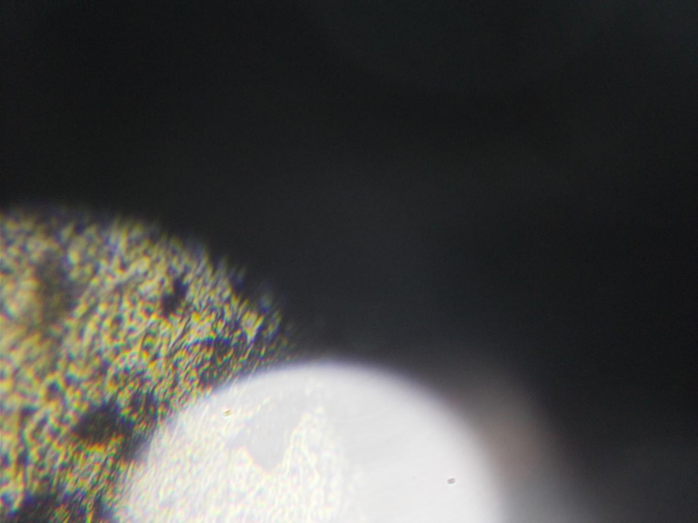
Lens wrapper. Stated 200 x. Actual c 130 x. (20.22.17)
Figure 7:
Lens wrapper. Stated 80 x. Actual c 75 x. Figure 8:

I had thought this was at 800 stated - I didn't initially think to write down the stated magnifications - but comparison with Figure 9 suggests it is instead a lower magnification, 200, rather than the same image taken further away from the microscope, although I might be wrong and will amend this as I look again at my observations. This was also taken earlier in November 2017 with less clarity than initial observations. Initially both the black line and the object below it disappeared as the focus was adjusted, appearing to be light distortion, and leaving the rest of the image relatively clear. With subsequent observations, both the light distortion and the rest of the image blurred as the focus was adjusted. The more translucent part of the image, in the lower part of the photo, is present whether or not the slide is present, is likely to be of part of the microscope itself and resembles more what I see with the Prinz. The same is true for Figure 9.
Figure 9:

This is taken at 800 stated, and by now the image at 800 had started to take on a more luminous look than at the beginning of the month. I will attempt by looking through the observations again when I have time - or my camera - to put the photos in chronological order.

[check observations] The same piece of Jaffacake wrapping (the top of the width of the film) at 200 x stated.
The same piece of wrapping at 80x stated.
80 x stated piece of film lit from below.

At 80x stated, a piece of film wrapping lit from below.

This was the top of a piece of light distortion sticking out from the edge of a piece of film wrapping, from memory and the size of the distortion, at 800 x stated.
Selected quotations
"According to Babeuf, Robespierre expected the population to be greatly reduced by the Terror, the war and the internal uprisings. He planned to achieve a redistribution of the land by the liquidation of the landowning class. Its members would be, if not killed off, forced to 'execute themselves' in time, and in their own interest." J.L. Talmon, The Origins of Totalitarian Democracy, 1952.
"If radical environmentalists were to invent a disease to bring human population back to sanity, it would probably be something like AIDS ... the possible benefits of this to the environment are staggering ... just as the Plague contributed to the demise of feudalism, AIDS has the potential to end industrialism." Christopher Manes, Green Rage: Radical Environmentalism and the Unmaking of Civilization, 1991.
“The world revealed by a microscope is not less wonderful than the world revealed by the telescope. … The wing of the fly is not only a thing of beauty, but is cleverly constructed on sound mechanical principles; the eye of a fly is not only beautifully made, but shows a knowledge of optics;”. 100 Peeps Through A Microscope, in ArthurMee (ed), The Children’s Encyclopedia, Volume 3.
"The case of sickle cell anemia and malaria is rather a special one, but beneficial evolutionary consequences of improved medical care may be quite common." John Maynard Smith, Eugenics and Utopia, 1965.
"It follows that as our ability to recognise heterozygotes increases, we could be led to sterilize almost the whole population on eugenic grounds, which is clearly absurd." John Maynard Smith, Eugenics and Utopia, 1965.
"Vide Ricardo's letter to Malthus of October 9, 1820: “No law can be laid down respecting quantity, but a tolerably correct one can be laid down respecting proportions. Every day I am more satisfied that the former is vain and delusive, and the latter only the true objects of the science.'" John Maynard Keynes, The General Theory of Employment, Interest and Money, 1936.
"We might conjecture that in the long run, if there is an upper bound on ability, we would eventually reach a society with the greatest equal liberty the members of which enjoy the greatest equal talent. But I shall not pursue this thought here." John Rawls, A Theory of Justice, 1972.
“An institution suitably located, constructed, organized, managed and personneled, to supply, scientifically, economically, efficiently and unhindered, all or any recognized part of the complex requirements for the prevention, diagnosis, and treatment of physical, mental, and the medical aspect of social ills; with functioning facilities for training new workers in the many special professional, technical and economic fields essential to the discharge of its proper functions; and with adequate contacts with physicians, other hospitals, medical schools and all accredited health agencies engaged in the better health program.” Quotation from the Council on Medical Education, entry under ‘hospital’ in Dorland’s Illustrated Medical Dictionary, 23rd Edition, 1962.
“Apparent magnification under the microscope.” Under ‘magnification’ in Dorland’s Illustrated Medical Dictionary, 1962.
“This sixth edition introduces widespread amendments, which reflect the ever-changing face of opthalmic practice. To keep abreast of changes in examination techniques, I have added an appendix of Multiple Choice Questions, as well as a short glossary, for both of which I am grateful to the help and patience of Dr Stuart Pinkerton, and which I hope will ease the passage of the hard-pressed student. Patrick Trevor Roper, 3 Park Square West, London NW1”. Patrick D. Trevor-Roper, Lecture Notes on Opthalmology, Sixth Edition, 1980.
“The great conquerors, from Alexander to Caesar, and from Caesar to Napoleon, influenced profoundly the lives of subsequent generations. But the total effect of this influence shrinks to insignificance, if compared to the entire transformation of human habits and human mentality produced by the long line of men of thought from Thales to the present day, men individually powerless, but ultimately the rulers of the world.” Alfred North Whitehead, Science and the Modern World, Lowell Lectures, 1925, 1967.
“This work [volume I, 827 pages] is intended for continuous reading, being designed as a course of study. In dealing with an enormous field of knowledge the editors have thought it best to omit consideration of subjects adequately treated elsewhere in accessible works. They have excluded from their survey, for example, the development of medicine, of architecture, and of certain other arts.” Charles Singer, E.J. Holmyard and A. R. Hall, A History of Technology, Volume I, 1958.
“A microscope using light will not magnify things clearly more than about 3,000 times, whilst an electron microscope can already magnify up to 300,000 times, and will probably do much better in future. … So far as I know, no photographs have yet been taken to show the detailed structure of human cells, but this will doubtless be done soon if it has not been done already.” J.B.S. Haldane, Science Advances, 1949.
“Executive order 8840, dated July 30, 1941, established the Office of Coordinator of Inter American Affairs within the Office for Emergency Management. The Coordinator took over the functions of the Office of Commercial and Cultural Relations Between the American Republics established by order of the Council of National Defense on August 16, 1940. The Office of the Coordinator of Inter American Affairs coordinates all activities of this country, designed to improve cultural and commercial relations among the nations of the Western Hemisphere. It operates both through governmental and private agencies in such fields as the arts and sciences, education, and travel, the radio, the press, and the cinema.” In US Government Monthly Catalog issued by the Superintendent of Documents, No 564, December 1941, page 1697. The significance of this may seem less obvious than that of some of the other quotes, but there are many suggestions that history has been re-written and whether or not this is an authentic record, the emphasis on ‘inter American affairs’, and lack of reference to Pearl Harbour, although troops in Washington on December 12th, as well as a reference elsewhere to optical instruments and a research paper, RPI, 1430, on ‘Reflection-Transmission relationships in sheet materials’ by Herbert F. Launer, 1941, suggests one avenue of research into the history of when and why disease was invented, although there are likely to be blind alleys.
“Two contrary laws seem to be wrestling with each other nowadays: the one, a law of blood and death, ever imagining new means of destruction and forcing nations to be constantly ready for the battlefield; the other, a law of peace, work and health, ever evolving new means of delivering man from the scourges which beset him. The one seeks violent conquests, the other the relief of humanity. The latter places one human life above any victory; while the former would sacrifice hundreds and thousands of lives …..” Louis Pasteur, 1888, quoted in Kenneth M. Smith, Beyond The Microscope, 1945.
“Now the war is over, I am beginning to start work again on these problems, though I cannot do much till University College is rebuilt. The Nazis have done such horrible things on the basis of their false theories of heredity that all work on this subject is inevitably suspect. But we can best counter these falsehoods by discovering the truth, and the truth is going to take the study of human, animal, and plant structure a whole stage beyond that which the microscope made possible.” J.B.S. Haldane, Science Advances, 1949.
Appendix 1: earlier observations with the Prinz, Sunagor MagnaScope and Apex Learner [draft report]
The first observations were from the Prinz 2801, made while assuming that what one saw when the microscope was illuminated from the mirror was all one could see with the microscope:
The lenses are the wrong way around: we would expect to see a smaller image where the first lens is larger than the smaller for the length of the tube whether or not the eye also leads us to expect a smaller image. With the Prinz 2801 microscope we do not appear to see anything beyond the microscope except as a pale ‘wash’ of the stain over a more complex image of something within the microscope, although at the moment I have no way of knowing if a different illumination would produce different findings. With the Sunagor MagnaScope and the Apex Learner we see beyond the microscope and magnification is compounded – ie, both lenses add to the size of the image - but the image we view is at significantly lower magnification than claimed, although there is less ambiguity as to what we are seeing with different objects using the Sunagor: at high magnification with the Apex Learner, as well as the blurred image of the stain we see a structure similar to that with the Prinz and likely to be of something within the microscope, although this conclusion requires corroboration. With none of the microscopes can we see clearly at more than 100 times, and perhaps less, the imprecision due to our not being able to determine precisely the width of the screen we are actually viewing nor to distinguish between light distortion and enlargement.
The Sunagor MagnaScope recently appeared to magnify a piece of film cut to 1 mm around 4-5 times with the microscope (ie, with the zoom added to the magnifying glass). Apparent clear magnification seems intuitively to be higher when we first look at the image but seems to be because we overestimate the size of the screen, since if we compare the actual size of individual letters to the size they appear to be on the screen, the magnification appears to be closer to four to five times. If one removes the funnel of the microscope away from the microscope, the image blurs to the point of invisibility very quickly – ie, within a few millimetres – which implies either that the limits of convexity have been reached or, given the shape of the funnel and the blurring rather than reduction, diffraction of light. Enlargement with the microscope - ie, with the magnifying glass and zoom - is considerably less – around one fortieth - than the 196 times (28x and 7x) claimed and less than would be needed to see a ‘cell’. Enlargement can be explained by partial re-reflection from the magnifying glass lens to the lower of the zoom lenses (see section on Reflection). The magnifying glass appears to magnify clearly at around 2 x . As I lift my eye from the magnifying glass, the image blurs and then turns upside down. The zoom also appears to magnify about 2x, although the effect is to produce an image of around the same size as the object, as if we were standing at the correct position to view it in focus. One interesting observation with the Sunagor MagnaScope is that when I removed my eye from the eyepiece of the zoom when it was attached to the magnifying lens while holding the funnel away from the eyepiece and straight in front of me , in other words when I would expect to see a blur, I could see a clear image of what was beyond the microscope on the edge of the eyepiece lens , as though the objects in front of us were first being reflected onto our own eye before being reflected onto the zoom. Although there might be other explanations, this would be consistent with what I observed in the science section of the Newark Museum, New Jersey, when there seemed no other mechanism by which the objects beyond the box could be observed on the screen on the nearside of it. On the other hand, I have not been able to get the same result during a recent observation with the Sunagor and so will need to repeat the observation under different conditions.
Since then I have estimated approximately 14 times using a tape measure (7mm visible on a screen estimated to be about 100 mm), which is closer to what I had originally thought, which was that the microscope magnified by about 12 times (rather than the 196 stated, unless of course magnification is thought of as area). This might indicate I have underestimated the width of the film when using the Apex Learner and so I will need to repeat the observations but also with another object, which of course I need to do anyway to support the claim that the image is a constructed one.
The first observation with the Apex Learner was done using a piece of bronze film – the ocular lens wrapping - cut to approximately 1 mm wide and assuming a screen diameter of about 60 mm with 10x ocular lenses and 80mm with 20x – the actual width is difficult to gauge and the inclination is to underestimate it so as to confirm the hypothesis of low magnification, but the assumptions are based on comparing the circle one sees and any line across it, such as the lash of the eye, with circles of different diameters and a tape measure Lighting was from below and above; the circle tends to appear larger if we have been looking at measured circles and then at the screen than if we take a break and then go to the screen before looking again at the circles. Subsequent observations were made : using a tape measure, assuming marks of about ¼ of a millimetre wide between the millimetres or a red thread of a similar width (ie, ¼ of a millimetre), and a page of writing, in both cases using lighting from above. Screens of 60 and 80 would represent about 6-12 and 8-16 x magnification of the objective lens tube depending upon where in the tube the lens is and assuming we are viewing the lens tube rather than the condenser or cylinder. The observations were done forgetting I could adjust the position of the slide, although this demonstrated the precision necessary even at fairly limited magnification, and allowed me to see magnification using the 20 x and 40 x, which I was unable to focus on when the film was under a slide casing and the objective lens lowered.
(1) With 10x ocular and 4x objective – ie, a stated magnification of 40 x – the piece of foil appeared to take up about 1/10 to1/4 of the screen or about magnification of 6 to 30 x, the difference appearing to do with the amount of light distortion; however it is positioned, however, the image is not blurred and not at all complex - ie, a wash of bronze; we can see a clear image if we lift the foil from the slide closer towards the aperture; then we can see a remarkably complex image of light and dark taking up 1/5 to 1/6 of the of the screen, a magnification of about 10 to 12 times.
(2) With 20 x ocular and 4 x objective – ie, a stated magnification of 80 times – the piece of foil appears to take up 1/3 to ½ of the screen but is not clear or complex; if we lift the foil, higher than with the 10 x ocular, we again see a complex image, this time taking up ¼ to 1/3 of the diameter of the screen , but clearer at ¼, or a magnification of about 20 x.
(3) With 10x ocular and 10 x objective – ie, a stated magnification of 100 x – the foil takes up ½ to a whole of the screen – ie, a magnification of 30 x to 60x, the difference appearing to be light distortion, and the foil appearing as a very pale bronze, the illumination apparently compensating for greater opacity (as with the 10x and 20x lenses in i and ii above); if we hold the foil half way between the slide and the plane aperture, we see the thread taking up about 2/3 the width of the screen, an apparent magnification of 40 x of a remarkably complex image, with a high degree of clarity, although some blurring and colouring at the edges.
(4) With 20x ocular and 10x objective – ie, a stated magnification of 200 x – the foil is now a darker shade of bronze, implying greater opacity, and appears to take up the whole of the screen; with the 10x objective, less of the screen appears to be visible at one time than with the 4x, as we would expect with greater convexity; the image is unclear, no more than an undifferentiated wash, although with the 20 ocular, the cornea of our own eye is more visible than with the 10x and we can see the beginning of a fixed structure, mainly a few dark spots; as one lifts the foil, it again comes into focus and covering a similar width of the screen, although more obvious than with the 10x ocular is that it is difficult to view the whole of the image in focus, although this might be to do with a slight folding in the foil, but also that it comes more into focus as we lift our eyes from the eyepiece, so that the apparent magnification decreases, the size of the screen appearing to be smaller; it therefore may be unlikely that we are obtaining more than 40x magnification.
(5) It is difficult to obtain consistent results at 4, 10, 10, and 20 and the observations need to be repeated.
(6) At 10x and 40x – ie, a stated magnification of 400 x – a complex image is revealed on the screen whether or not anything is present; the corneas of the eye are present above it; and the foil appears as a pale film over the image; I was able to observe the structure of the foil when I lifted it this time the screen appearing about 50 to 60 mm and the foil width being about double that, which would imply a magnification of around 100 x; it is still complex and reasonably clear, although judgement is perhaps made more difficult by the fact that one is not looking at familiar shapes; also it is difficult to hold the foil in the same place, which in itself also suggests quite high magnification.
(7) At 20 and 40 x – ie, a stated magnification of 800 x – I could still find a relatively clear complex image of the foil when lifted it from the slide and placed quite close to the lens, with my eye still quite close to the eyepiece, but with not significantly greater enlargement than with the 10x – ie, the width of the foil did not have to travel across 10 to 20 screens before it was no longer visible; the image was darker, and fairly difficult to focus on, especially since the fixed image and corneas were also present, and the individual parts seemed larger rather than that the object was more complex than at lower magnifications, although this needs to be established with more observations; but it was still fairly clear, at what looked like a magnification of at least 100.
Further observations to those above and those following the introduction will be done with objects where it is easier to establish the extent to which clarity decreases with higher magnifications than with those that will necessary resemble the structure of living tissue more closely than that of the foil, although it also needs to be established whether more complex structures than those observed in the foil would be revealed further away from the lens with different objects, which seems unlikely. The magnifications estimated were based on measurement of length. If magnification is taken to be measurement of area, then I have in fact underestimated apparent magnification, which would be greater than 800 times.
Further observations:
(1) With 10x ocular and 4x objective lenses, magnification appears to be about 40 x using a piece of tape or a thread because the line or thread is visible on about one third of the screen; however the line is very blurred and paler than the original, nor is any ‘structure’ revealed.
(2) However, if I place a piece of translucent bronze foil about 1 mm thick under the lens, it seems to take up about half the screen, implying nearer to 20x magnification, so that some of the magnification one saw of the red line might have been distortion.
(3) Lit from below, it seems to take up less of the screen, one third or less (if one tries to see the screen at its widest, ie, with one’s eye to the eyepiece, the light is very bright), but with less distortion, ie, a clearer outline, and implying magnification of around 13 times.
(4) With 10x and 10x lenses, and lit from above, the thread appears to cover the screen but not much more of it, implying a magnification of near to 100 but with considerable blurring and with possible overestimation of the width of the screen in the cylinder; however, the structure of the thread becomes clearer at lower magnification closer to the aperture, ie, with small threads at an angle from the main thread.
Additional points: With 4x, 10x and 40x objective lens tubes alone – ie, with the eyepiece removed and when peering down the cylinder - we appear to get disproportionate magnification, with about 20mm of the tape being visible with the 40x and only 3 mm with the 4 x and 4 mm with the 10 x. This would seem to be, first, because the light above the lens illuminates a larger area than that of the circle underneath the opaque tape. The fact that one appears then to see a larger area with the 40 x may be that the aperture in fact acts as a condenser whereas there is more diffuse illumination onto the larger apertures so that we have only partial reflection onto the lenses. On the other hand, there may be some interplay between the lenses, implied also by the 4x, 10x and 40x, but further thought and observation is needed. Although 4x, 10x, and 40x lenses might have been chosen to suggest compounding, in whichever direction, of magnification within the objective lens system, I have not yet thought through how this might be or not be achieved. The 20x ocular lens is shorter than the 10x and has a smaller lens. With the 10x ocular lens alone, ie, attempting to use it with a magnifying glass, there is magnification probably closer to 5x than 10x, but the object is blurred and upside down. With the 20x ocular lens, there is relatively clear low magnification if we have our eye away from the lens but as we attempt to increase magnification by moving our eye away further there is blurring and we see the image move upside down. It does look as though one might see the lens of one’s own eye in an upper layer since the image appears to change with different eyes although that might be that I am much more short sighted in one eye than the other. One appears to be viewing something projected beyond the ocular lens because I cannot see the pink ‘wash’ from the slide with the left eye. With lighting from below only I can see the pink wash of the stain on the slide with some difference in shade of pink with the different objective lens tube magnifications consistent with lower and higher magnifications. With the addition of a second lens, we might therefore be expected to still distinguish areas of white and pink with the lower magnification lenses but an undifferentiated lighter pink with the higher magnification where we have many lines close together, which is somewhat consistent with the point above, although better conclusions could be drawn with more observations or perhaps different eyepiece lenses.
Second observations with the Apex Learner:
These were with the 10x eyepiece lens only and using the same piece of foil as the previous month, the width of which is difficult to measure with accuracy because it is not of exactly the same width, is folded in the middle, and perception of whether the semi translucent foil covers one or two millimetres can be difficult to determine. However, in basing on what follows on the assumption that the foil is only 1 millimetre wide, I will not be underestimating magnification because it can seem as much as 2 mm in places. This time the piece of foil was on the slide and with a slide casing fastened on top, although it would have been easier to manipulate had it been placed horizontally rather than vertically, which I will do for the next observation with a more carefully cut, if I can, piece of foil. I did not measure, because of the manipulation problem, the amount of the stage visible.
(1) With 10x eyepiece and 4 x objective lens: the screen this time appeared to be 100 mm, although as last time, appeared of a smaller diameter when I went back to it. The width of the foil seemed to be about 30 mm, by comparing it with the length on the tape measure rather than relative to the screen. An illumination of 30 is not much lower than the 40 stated.
(2) With the 10x and 10x eyepiece and objective lenses, the screen this time looked somewhat smaller and then 100 mm again and this time it was easier to estimate magnification by comparing it to the diameter of the screen: the width of the ribbon appear to be about the width of the screen, which would be consistent with a 100 mm length diameter. Also, there was better resolution, in the sense that it was easier to distinguish between parts in an object within the ribbon, although they could not yet be seen clearly. By easier to see, I mean that I had more of an impression of lots of parts in a small flower-like object but it is easier to distinguish and count with the 10 x objective.
(3) With the 10x and 40x eyepiece and objective lenses, I judged that the ribbon covered about 2.5 times the width of the screen, which again I estimated to be about 100 mm, which would mean a magnification of 250 rather than 400,. Given the greater discrepancy here, I will repeat the observation with a ribbon placed horizontally. However, of interest was that the image was clear and small parts were easier to see, again in the sense of attempting to count the number. While the smallest parts were discernible but not entirely clear there was a clearer outline of the flower-like object than with the 10x eyepiece and so in this sense there is better resolution with the higher magnification. However, the extent of the magnification will need to be determined with another observation.
Further observations to those above and those following the introduction will be done with objects where it is easier to establish the extent to which clarity decreases with higher magnifications than with those that will necessary resemble the structure of living tissue more closely than that of the foil, although it also needs to be established whether more complex structures than those observed in the foil would be revealed further away from the lens with different objects, which seems unlikely.
Appendix 2: the diameter of the aperture on the Prinz 2801
In theory, a convex lens – eg, one resembling a glass bead in shape - might enlarge an infinitesimally small speck to the width of the aperture or lens; whatever the difficulties and consequent distortion and weakening of the image, the size of the apertures are not correct, even allowing for approximation of measurement. The following measurements are approximate, it being difficult to measure exactly because of the obstruction of the microscope stage. They were also based on the assumption that the aperture would be too small rather than too large and so the calculations were carried out at the lowest point possible for the lens to bel.
Magnification from the objective lenses of the |Prinz 2801 is said to be 60 x, 30 x and 10 x.
Actual aperture diameters: 3 mm (60 x); 1 mm (30 x); 2 mm (10 x) : for the same length of objective lens tube we would expect a larger diameter with a lower magnification because the angle of reflection is smaller, assuming the diameter of the lens is the same; in fact, the aperture for 30 x is smaller, not larger; the aperture for 10 x is larger than for 30 x but the tube is shorter and the aperture size is smaller than for 60 x.
Below I have calculated the size the aperture should be assuming: light from a convex lens of the same widths and in different positions in the microscope (i) to (x) and a ratio of reflection of 60, 30 and 10 times from slide to an 8 mm diameter lens in the objective lens tube (xi to xiii); assumed sizes based on measurement rather than calculation. Since the initial calculations are based on simple ratios only, the last three only need be read.
(i) assuming lens 8 mm diameter, 23.5 mm from slide casing, aperture 5.5 mm from lens (60 x and 30x): aperture size: (5.5 divided by 23.5) x 8 = 1.87 mm (to 2 dp);
(ii) assuming lens 8 mm diameter, 23.5 mm from slide casing, aperture 11.5 mm from slide casing (10x): aperture size: (11.5 divided by 23.5) x 8 = 3.91 mm (to 2 dp);
(iii) assuming lens 8 mm diameter, 13.5 mm from slide casing, aperture 5.5 mm from slide casing (60x and 30 x): aperture size: (5.5 divided by 13.5) x 8 = 3.24 (to 2 dp);
(iv) assuming lens 8 mm diameter, 13.5 mm from slide casing, aperture 11.5 mm from slide casing (10 x): aperture size: (11.5 divided by 13.5) x 8 = 6.81 (to 2 dp);
(v) assuming lens 20 mm diameter at top of lens tube and at height of condenser; 23.5 mm f rom slide casing; aperture 5.5 mm from slide casing (60x and 30 x): aperture size: (5.5 divided by 23.5) x 20 = 4.68 mm (to 2 dp);
(vi) assuming lens 20 mm diameter at top of lens tube and at height of condenser; 23.5 mm from slide casing; aperture 11.5 mm from slide casing (60x and 30x): aperture size: (11.5 divided by 23.5) x 20 = 9.79 mm (to 2 dp);
(vii) assuming lens 50 mm diameter, ie width of condenser (presenting problems of viewing directly and reflection if lit by mirror); 27.5 mm from slide casing; aperture 5.5 mm from slide casing (60x and 30 x); aperture size: (5.5 divided by 27.5) x 50 = 10;
(viii) assuming lens 50 mm diameter, ie width of condenser ; 27.5 mm from slide casing ; aperture 11.5 mm from slide casing (10 x); aperture size: (11.5 divided by 27.5) x 50 = 20.91 mm (to 2 dp);
(ix) assuming lens 30 mm diameter, ie width of lens tube (assuming direct viewing of screen); 53.5 mm from slide casing; aperture 5.5 mm from slide casing (60 x and 30 x); aperture size: (5.5 divided by 53.5) x 30 = 3.08 mm (to 2 dp);
(x) assuming lens 30 mm diameter, ie width of lens tube; 53.5 mm from slide casing; aperture 11.5 mm from slide casing (10 x); aperture size: (11.5 divided by 53.5) x 30 = 6.45 mm (to 2 dp);
(xi) assuming magnification from 0.133 mm to 8 mm lens at height of condenser (60 x): lens at 23.5 from slide casing; aperture at 5.5 from slide casing: aperture size: 0.133 x (5.5 divided by 23.5) x 8 = 0.25 mm (to 2 dp);
(xii) assuming magnification from 0.27 mm to 8 mm lens at height of condenser (30 x) : lens at 23.5 from slide casing; aperture at 5.5 from slide casing: aperture size: 0.27 x (5.5 divided by 23.5) x 8 = 0.51 mm (to 2 dp);
(xiii) assuming magnification from 0.8 mm to 8 mm lens at height of condenser (10 x): lens at 23.5 mm from slide casing; aperture at 11.5 from slide casing: 0.8 x (11.5 divided by 23.5) x 8 = 3.91 mm (to 2 dp).
If the aperture should be either closer to the lens or larger, as is the case with the higher magnifications, this suggests one of two things: (i) we would not see the object because it is beyond the focal point of the lens; (ii) the image would be reflected onto a part of the lens only so that, given the constriction of the lens tube and cylinder, the maximum possible enlargement would not be obtained. Crowding of light around the entrance of the aperture would also undermine the function of the condenser, which is to concentrate the flow of light.I
If it should be either further from the lens or smaller, as is the case with 10x, then this would also not be optimal because the light would not be guided sufficiently, resulting in a less clear image.
Bibliography
Britannica Concise Encyclopaedia (Encyclopaedia Britannica, Inc, 2002)
Breithaupt, Jim, Understanding Physics For Advanced Level, Second Edition. (Cheltenham, STP)
Chamot, Emile Monnin, and Clyde Walter Mason, Handbook of Chemical Microscopy, Volume I (New York, John Wiley & Sons, 1931)
Dawkins, M.J.R. and K.R. Rees, A Biochemical Approach to Pathology (Edward Arnold, London, 1959)
Dorland's Illustrated Medical Dictionary, Twenty Third Edition (Philadelphia and London, W. B. Saunders, 1957)
El-Kareh. A.B. & El-Kareh, J.C.J., Electron Beams, Lenses, and Optics, Volume 1 (New York, Academic Press, 1970)
Gosse. P.H, F, R.S., Evenings at the Microscope (Society for Promoting Christian Knowledge, London, 1877)
Haldane, J.B.S., Science Advances (Woking, Unwin Brothers, 1949)
Harris, Robert, Cancer: The Nature of the Problem (London, Reading and Fakenham, Cox and Wyman Ltd, 1962)
Hecht, Eugene, Schaum's Outline of College Physics, 11th Edition (Schaum's Outlines, McGraw Hill, 2012)
The Hutchinson Encyclopaedia (Oxford, Helicon Publishing, 1994)
Massachusetts Historical Society, Adams, Henry, The Education of Henry Adams, An Autobiography (Boston and New York, Houghton Mifflin Company, The Riverside Press, Cambridge, 1918).
Maynard Smith, John, 'Eugenics and Utopia', in Frank E. Manuel, Utopias and Utopian Thought (Boston, Beacon Press, 1966)
Mee, Arthur (ed), The Children’s Encyclopedia, volume 3 (London, The Educational Book Company Limited, no publication date).
Meinig, George E., Root Canal Cover Up (Ojai, California, Bion Publishing, 1994)
Orrock, Louise, On HIV and Aids, Taxila Institute (institute.iqmind.org/on-hiv-and-aids-by-louise-orrock), June 8, 2015
Orrock, Louise, On Cancer, Taxila Institute (institute.iqmind.org-on-cancer-by-louise-orrock), June 8, 2015
Oxford Dictionary of Science, Sixth Edition (Oxford, OUP, 2010)
Peckham, Michelle, What is Histology, Faculty of Biological Sciences (http://histology.leeds.ac.uk)
Poole, Lynn and Gray, Scientists Who Work With Cameras (New York, Dodd, Mead & Company, 1965)
PubMed, US National Library of Medicine and National Institutes of Health, (www.ncbi.nim.nih.gov/pubmed)
Singer, Charles, E.J. Holmyard and A.R. Hall, A History of Technology, Volume I, From Earty Times to Fall of Ancient Empires (Oxford, Clarendon Press, 1958).
Smith, Kenneth M. Smith, F.R.S. (Harmondsworth, Penguin Books, 1945)
Trevor-Roper, Patrick D, Lecture Notes on Opthamology, Sixth Edition (Oxford, Blackwell Scientific Publications, 1983)
US Government Monthly Catalog, Superintendent of Documents, No 564, 1941.
Whitehead, Alfred North, Science and the Modern World, Lowell Lectures, 1925 (New York, The Free Press, 1967)
Wikipedia (on diffraction; although a frequent user of Wikipedia, I have so far relied on books to provide standard accounts of magnification and the microscope).
Louise Orrock
© 2017

Does the microscope achieve high enough magnification to view the ‘cell’?
“So the physicists have produced a microscope known as the Electron Microscope which does not use light rays at all but operates with cathode rays. Instead of a beam of light it employs a stream of electrons, and electromagnets instead of lenses.” Kenneth M. Smith, Beyond the Microscope, 1945.
“[I]n 1904, Arthur Balfour announced on the part of British science that the human race without exception had lived and died in a world of illusion until the last year of the century.” The Massachusetts Historical Society, The Education of Henry Adams, An Autobiography, September 1918.
Introduction
Although cancer was apparently diagnosed before the invention of the electron microscope, diagnosis since at least the 1960s has relied upon it. I have had no access to an electron microscope but was initially sceptical that the traditional light and lens microscope viewed what was beyond the lens or, if it did, that it could achieve anything like the higher magnifications claimed. I then saw that high magnification was apparently possible with a traditional microscope, as much as 600x with the Apex Learner. However, because of distortions at low magnification with magnifying lenses I thought that the light microscope must then be an electron microscope, neither microscope in the sense of replacing light with beams of electrons, but because they must allow remote accessing and the construction of an image unless, in the case of the light microscope, a kaleidoscopic effect is produced mechanically within the microscope. However, I have not yet ruled out the possibility that I am seeing a significantly enlarged image of the object itself rather than a reconstruction and that the complexity and focus is the result of better illumination, as is implied by the name ‘light’ microscope.
However, the fact that the magnifications are not those claimed by the manufacturer, that some of the complexity and magnification appears, in fact, to be light distortion and that I have not yet obtained consistency in terms of the size of the object as a whole and its individual parts, that we are unable to measure enlargement precisely because we are estimating the size of the screen and the extent to which the object exceeds it, that the increase in complexity at high magnification seems less suggestive of underlying structure if we examine the object with the eye alone under strong light, that the measurement of magnification for diagnostic purposes is in length only so that the highest magnification the light microscope is capable of is nevertheless insufficient to diagnose disease, the implausibility of electron beams and other mistakes in explanations of magnification, including the fact that we are not viewing the object directly with lens magnification but, from my observations, are seeing a reflected image, has encouraged me to think I am right that microscopes do not view cells, even if the conclusion may be less obvious than if it could be demonstrated that the light microscope were not viewing the object on the slide or enlarging much more than can two magnifying lenses.
The following is based on observations using the Prinz 2801 (a standard two-lens light microscope), the Apex Learner (some modification to the standard shape), and the Sunagor MagnaScope (a two-part microscope consisting of a zoom and magnifier with a funnel to direct light). Each of these are light microscopes that depend on battery or mains, mirror or natural lighting and magnifying lenses to achieve enlargement. Until recently I had underestimated what could be seen using the light microscope because I had depended only upon the Prinz 2801 and Sunagor MagnaScope, both of which relied on natural lighting after damage, one of which could apparently not see beyond the microscope, even if it showed quite complicated and ‘cell’ like structures on the screen, and the other which could, but with magnification of what looks like only around 15 times in length, but with only natural lighting, the light unit having snapped off soon after I acquired it.
However, the Apex Learner, which is illuminated either from above or below the slide, achieves surprisingly high magnifications. With the Apex Learner I appear to see a two-millimetre wide piece of transparent bronze film – the ocular lens wrappings - magnified so far by as much as 600 times –360,000 times if we think of magnification instead in terms of area, although pathologists do not. However, it is impossible to assess the actual magnification since we are judging the width of the screen we see within the cylinder rather than an image projected onto a screen (and for this reason too it can be difficult to obtain consistent results). However, magnification appears to reveal structures that cannot be seen by the eye alone, although with the object observed (the bronze-coloured wrapper) these appear to be a pattern of opaque and translucent (reversed if we light from below rather than above) rather than an underlying structure.
When I obtained the higher magnifications I did not doubt that microscopes did not diagnose disease, which I have been certain of since guessing in late 2012, but was not sure how to account for it. The telescope contains mirrors, but whether or not there are mirrors inside the microscope – and I have so far been unable to take any of them apart - these would not, from observation of what one sees , for example, if one places a mirror between two magnifying glasses, add significantly to enlargement Instead, I wondered first why it would have been necessary to have invented the implausible-sounding electron microscope rather than continue to use the light microscope for diagnosis of disease unless it were to discourage amateurs and then whether what was being implied by the name was not in fact beams of electrons providing illumination but that all microscopes are electron microscopes in the sense that they resemble other electronic communications devices: the outline of the image or the object or stain itself is detected, enlarged and filled with a constructed complex structure.
The microscope as a diagnostic instrument
The sections following observations from the Apex Learner are sceptical with respect to traditional accounts of magnification but have been influenced by hints in these as well as in other non-fiction and literature. There appear to be mistakes in standard accounts of magnification. For instance, we are viewing a reflected image rather than the object itself, so that what is actually happening within the lens is re-reflection, determined by the character of the lens and illumination. The highest magnifications I have obtained are also lower than those claimed by the manufacturer, even if surprisingly high. The highest magnification possible with the light microscope, according to the department of biological sciences at Leeds University is 1400 times, less than that needed for cell pathology. However, given that this is almost 2 million times if one thinks of enlargement in terms of area, one wonders if the definition of magnification in terms of length was not intended to prevent self-analysis of cells.
Accounts of the electron microscope, on the other hand, assert that higher magnification and resolution, or clarity, can be obtained with electron beams and magnets, with magnifications of as much as 7 million times. [encyclopedia of biology] The mechanisms chosen, electron beams and magnetic coils, may be intended to undermine criticism of diagnosis, as might also the subject of light waves, or to encourage it, although the name may also be chosen to imply that what we understand to be a new kind of microscope may instead be remote accessing and construction of the image.
Dorland’s Medical Dictionary defines magnification as “apparent magnification under the microscope”. We can see very small objects – such as mould spores – with the eye alone, while the eye itself can achieve relatively high magnification: the structure of the cornea of the eye is revealed when we squint into light. Also, with limited magnification, especially but not only when using a zoom lens, we can have the impression of significant magnification when, if we actually compare the size of what we are viewing with the size of the object itself, we realise there is little or no actual increase in size: the illusion is created by viewing an object that is further away. Also, with a magnifying glass we may notice that it is the eye itself that is doing the magnification: for example, if we place a lens directly above an object on a page and then hold the lens closer to our eyes, the object will appear larger than when viewed without the lens but so may the lens itself, ie, the object may occupy the same proportion of the lens, although usually it will occupy somewhat more . Nevertheless, the light microscope does also appear to achieve surprisingly high magnification when we compare it to a camera that blurs at limited magnification (from observation, as little as 2 to 5 times, depending upon what we are viewing and certainly more than is possible with the eye alone. Dorland’s phrase may be referring to the fact that it is impossible for the microscopist to measure magnification in the traditional microscope - without taking it apart, which I have not been able to do with the Prinz or the Apex Learner - because we do not have certainty as to the width of the screen we are viewing, whatever the stated or calculated (ie, taking into account convexity, refraction, positions and size of lenses) magnification of the ocular lenses and the width of the objective lens tubes. Or he may be referring to the character of the electron microscope. Although I have as yet no knowledge of electronics, whether or not the magnetic coil detects the object or its reflection, while any subsequent increase in size is enlargement whether or not it is a reconstruction, the complexity is added, presumably as a result of external accessing and manipulation rather than from inside the microscope, so that we are therefore not viewing an enlarged image of the object on the screen. This would be consistent with the fact that we appear only to achieve relatively limited clear enlargement with light projection and magnifying lenses.
All enlargement using light projection or lenses results in loss of clarity. This is self evident: although we can bring an object into focus that is otherwise too far away or too close to bring clearly, any enlargement will weaken in the sense of blurring the image. With light projection, we may be able to view a slide significantly beyond its size but the image will be less clear than the object at the same size and blurring and loss of visibility occur as the size increases or because of insufficient light at greater distances. The image is dissipated, or becomes less clear, with projection because, as with any enlargement, the space between outlines increases but is also distorted because the angle at which the object is projected is smaller nearer the centre. It disappears when the lines that define objects have disappeared or when the light can no longer reach far enough or is too diffuse. When using a camera we can bring an object closer but often not enlarge without loss of clarity or, much beyond 2 – 5 x (depending on what we are viewing), identify objects. A zoom may allow us to see an object in the distance relatively clearly. However, even without enlargement beyond the actual size, we will see a less clear image than the original because the effect of a glass lens is to reduce rather than increase clarity, which we can observe if we were to move closer to the object and view it at normal focal length, or if we compare the image obtained with a zoom with that obtained if we improve focus by cupping our hands in front of our eyes.
“1. (in optics) A defect in the image formed by a lens or curved mirror.” Entry for ‘abberration’ in Oxford Dictionary of Science, page 1.
Whereas with slide projection, although there is an increasing loss of clarity, the image we see increases in size as we move the object from the slide, with a magnifying lens (or microscope) we are not attempting to project an image beyond its location and there is not an indefinite advantage in placing the object at an increasing distance from the lens although magnification also increases as we remove our eye from the lens as though projection of the object is continuing beyond the lens, although as when we think the objects in a mirror change when we look at it from a different angle but they do not, so the reflected image remains the same whatever our perception of it. This needs further consideration but can in part be explained by the fact that although the lens appears to remain the same size the size of the screen with a larger number of objects on it appears much larger when our eye is against the eyepiece, although not entirely since the actual size of the individual object on the screen appears greater when viewed from a distance. It is as though the convexity of the eye itself were resulting in a smaller area being reflected onto the eye, the eye itself mimicking the action of the lens and compounding magnification, but this will need further consideration.
With lens magnification, we get blurring, loss of visibility and distortions with enlargement, and the object then becomes smaller as the effect of convexity reverses and recedes. With convex lenses, the distorting effect is also greater than with enlargement from projection alone because the angle of enlargement is determined by curvature as well as illumination. We can see the distorting effect of lenses if we look at ourselves in a slightly curved mirror. Reflection - which the next section explains is what is actually taking place with lens magnification - is usually onto a three dimensional object (the lens) whose surface is different from that of the object being viewed, unless it is a similarly shaped sphere, the distorting effect being greater with greater magnification. The three dimensional nature of the lens also increases relative flattening at the edges. Other distortions include: uneven magnification and distortion of shape, light distortion, the image turning upside down, objects being displaced from left to right or vice versa, multiple images, increasing opacity, a tunnelling effect, and will be considered after discussion of what is occurring when lens magnification takes place, after a summary of observations from the Apex Learner.
“If well corrected lenses are used, the magnifying power of the microscope should be at least that necessary to reveal the finest details resolvable by the objective. For the normal eye, this is equivalent to about 500 to 700 times the numerical aperture of the objective.” [1931]
Initial observations with the Apex Learner can be viewed in the Appendix but what follows are more recent observations where I obtained clear images approaching the magnifications claimed. My aim has been to establish the extent of magnification, although I intend once I have obtained consistent results to turn to the character of the image. Because one is estimating the size of the screen and objects on it and because of the difficulty of manipulating the image to then measure with accuracy the size of the object (a piece of bronze-coloured translucent film – the wrapping for the lenses - cut first to approximately 1 mm in width and then to 2 mm and placed under a slide casing), I have repeated the observations in an attempt to obtain consistency and to avoid the temptation to underestimate magnification and record true results.
(i) First, carrying out the observation with a piece of bronze translucent film placed horizontally on the slide, and cut fairly consistently to about 2 mm in width, the size of the width of the foil on a screen appearing to be about 100 mm appeared to be 50 mm, 120 mm and 450 for the three objective lens tubes with a 10x eyepiece lens, supporting magnification of 25x, 60x, and 225x. Manipulation was difficult because the image appeared less differentiated although no less complex than with a previous piece of film (see Appendix 1). Nevertheless, we see within the ribbon thousands of tiny parts, if not completely clearly, resembling something like the map of a city. Of interest in this observation was that I noticed that with illumination from below one saw the image in negative: which is consistent with the structure reflecting patterns of light and opaque. Magnification of 25x, 60x and 225x differed from the stated magnification of 40x, 100x and 400x, and further observations will be undertaken to establish whether the variance is real.
(ii) Carrying out the observation with the same piece of film - 2mm and the 20x eyepiece, the width of the screen appeared to be about 150 mm rather than 200 mm, but it was difficult to make the judgement. With the 4x objective lens tube , the width of the ribbon appeared to be at least 150, which would indicate a magnification of 75x rather than 80 x, an insignificant variance given the difficulty of estimating the width of the screen. With the 10 x objective lens, the width of the piece of film appeared to be about 300 mm, which would indicate a magnification of 150x. With the 40x objective lens, it was not possible to position the lens tube so that the film could be viewed in focus. Instead, I cut another piece of film and placed that on the stage without a slide or casing. Then the width of the magnified object did appear to be about 4x that of the same object (ie, a part of the film, given the difficulty of manipulating and keeping in focus the ribbon when not in a casing – and whether or not the apparently shifting image was or not the result of any sort of external manipulation, this was when I first thought that ‘electron’ microscope might be code for remote viewing of all microscopes). This would indicate a magnification of at least 600x (rather than 800 times). At this highest magnification we see a similar and remarkably complex image with the thousands or tens or hundreds of thousands (repeated observation may establish which) small parts, mostly roundish, some more rectangular, as well as wider lines, larger than at lower magnifications but not obviously otherwise clearer or more complex (apart from the more rectangular shapes being more visible than at lower magnifications) or more numerous. The variance between the magnification obtained with the Apex Learner and what I had seen with magnifying glasses seemed hard to account for. The image is also remarkably still at high magnification. This has led me to believe that although the light and lens are able to detect the shape of the object, the enlargement of the outline is a reconstructed one and the complexity added, so that the Apex Learner must be an ‘electron’ microscope.
(iii) Second, carrying out the observation with a piece of Jaffa cake wrapping 2 mm wide [added subsequently from memory so that I have changed the estimation of magnification that follows; I lost the two bits of film I was working with but think the biscuit wrapping was not 4 mm, which the initial estimation suggested], I found magnification of approximately 2-400x in length or 4-16,000 times in area, which is said to be the magnification needed to view the HIV cell; this was achieved with the 10x ocular lens and 40x objective lens and so may have reached the 400 x claimed. I estimated the screen width to be about 80 mm and the piece of foil to be about 5 times the width of the screen. During this observation I was less convinced than in the previous observation that there was either remote manipulation or even a kaleidoscopic internal adjustment of the image, even though the image was not entirely clear as I moved it. Nor did the stillness of the image seem as suspicious as when I had carried out the observations with the lens wrapper. This then seemed a remarkably clear image given the magnification, even if the increase in magnification of individual structures seemed more apparent than real in terms of the actual increase in length as opposed to area. At 4x I saw the foil cover most of the screen so that the stated increase from 40x to 400x was in fact by about five rather than one hundred times. If I hold the wrapper up to the light I can just see the edge of the wrapping - where I cut it. I can also see cracks in the wrapping and what look like dust particles that may account for some of the complexity. Comparing this with what one sees with a magnifying glass of 1.5 or 2. I can start to see, especially with strong illumination, that the complex structure is discernible, the lens wrapper particularly looking something like a layer of skin. While on the one hand it does not look like, for instance, the creases one can see with the eye alone have been magnified, say, 400 times, I cannot yet say with certainty that I am overestimating the size of the screen or that the complexity is constructed or reproduced, particularly where the width of the foil covers less than the screen. What I can say is that the magnifications claimed are fairly inaccurate, whether length or area, and that this seems odd, given that it does seem to achieve high magnification. and that the resemblance of the image under the microscope to what one sees when holding the film up to strong light may support significant enlargement but not the revelation of an underlying structure.
(iv) Repeating the observation again with the biscuit wrapper and the 20x eyepiece lens, what looked like a 2 mm long crack in the piece of wrapper was magnified to what looked like 40 mm with the 20 x lens and 4x objective (20x) but assuming a somewhat smaller - 60-80 rather than 80-100 than with the 10x lens - rather than larger screen than with the lower strength ocular lens. With the 10 x objective, the same crack was visible bus appeared to take up more than the screen, about 100. The length of the crack appeared to increase around 1.5 times, implying a magnification of between 30x (assuming the lower magnification was correct) and 50x. This is less than the 80 and 200x claimed by the manufacturer, although a lot more if the magnification were based on area, which it seems to be with the Sunagor MagnaScope. Although the stillness of the image is compatible with our seeing the object rather than a construction, neither on the other hand at clear magnification of 50x or 2500 x are we seeing any moving parts. With the 40x lens, interestingly, one can see that the widened edge of the wrapping is, in fact, distortion that, depending on the position of the lens, can be present or absent without loss of clarity of the rest of the image. At this magnification as with the 10x and 40x, what I take to be imperfections and particles on the eyepiece lens, are visible. I am less certain than I am with magnifying lenses that I am seeing a reflected image. However, whether or not the apparent difference between the glasses and the microscope is better lighting, notably in the case of the Apex Learner, from the round shaped light below the microscope stage, although we see more clearly and perhaps in more detail with the stronger of the two lights, the size of the outline is the same whichever of the two lights is used and also with natural lighting, where, however the detail is more difficult to see, which does not support the idea that we are seeing the underlying structure of the cell with the microscope. It also seems unlikely that however bright and guided the lighting that we would be able to see through the cylinder beyond the lenses rather than that the image is somehow captured in the lenses themselves. If it is thought unlikely that we would be able to see so clearly within a lens, the magnified structures or imperfections in the eyepiece lens are clear when the light is on and there is no object present, as is the image we see in a mirror. However, it does seem most likely that better lighting is responsible for the greater complexity rather than that there is an added construction. However, in the next observation I will look more carefully at light distortion - which at the end of the last observation I saw could produce an apparently clear image of a widened edge of the wrapping but which disappeared as I adjusted the focus so that the edge was not all that much wider than when viewed with the eye. I will also try and establish with more certainty that there is no kaleidoscopic effect, or reproduction of parts of the image within the screen.
(v) Repeating the observation with the 20x eyepiece lens and 40 x objective and using a piece of biscuit film wrapping in order to determine the extent of magnification only (ie, less concerned with the structure of the image), I found that the individual parts do not seem to be magnified much beyond 40x. This does not mean that the eyepiece lens is faulty because there is apparently magnification of more than 40x of the width of the film and because one can see compounding by comparing the image of an object with both lenses and by simply looking into the cylinder with no eyepiece lens present. I was surprised by the length of time it took for me to find an image even despite the difficulty of manipulating the film without it being on a slide under a slide case. What I have also noticed before but had not added here is the difficulty at times of distinguishing between the enlarged object and the underlying fixed image on the screen as well as sometimes the cornea of one’s own eye that is also present, although unlike with the Prinz 2801 I am, with the correct focus, obviously seeing a new image, which appears to be that of the object underneath the lens. I have also not yet become adept at manipulating the object lit from below (which it has to be at higher magnifications) so that when moved to the left it appears to move to the right. However, manipulating it by hand - ie, without a slide present - also means that it may be nearer or further from the lens and light so that the size of the object may vary as one moves it, as for instance, the object can disappear when the lens is closest to the microscope stage and also appears to shift significantly as I adjust the focus. However, manipulating it by hand is a problem at the highest magnification with this microscope only.
(vi) In a further observation with a 1 mm piece of the orange-coloured film and the 20x eyepiece lens, an object within the image appeared at about 30 mm on a 120 mm screen (30x) with the 40x and 10 to 15 mm with the 10 x (10x - 15 x)and about 5 mm with the 4 x (5x). However, the object disappears relative to the image as a whole - for instance, one sees a thick and complex edge of the ribbon disappear to a virtually invisible one - as one adjusts the focus, most clearly with the 40x lens, and in fact so do many of the small objects within the ribbon of film, not so much, as we can see with the 40x lens, because of loss of focus but because they are ‘light spots’ that disappear or come into view as one turns the focal adjustment. On a repeated observation it seemed more as though the spots or objects disappeared as I turned the focal adjustment as the object got larger or smaller but even so looking more like one sees in a kaleidoscope than the blurring you get with a camera. I was not able to estimate the width of the object this time because of the problem of manipulation and seeing where and there which width of the ribbon I was viewing.
(vii) Going back to the 40x and 20x the next day, the image was less clear in the sense that the eyepiece lens looked more opaque even after dusting. This is more in line with what the manufacturer also reassured me yesterday, which was that the image would not be clear at 40x and 20x. (They also told me the product was intended at primary school children, whereas I thought I had read it was what pathology students trained on; it makes no difference except perhaps to discredit what I am doing in the eyes of those entirely unfamiliar with the microscope.) Since the light seemed also to be worse for the eye, I did not try again the same day. I was, however, pleased that a mark that I thought had got onto the condenser and might have been confused with an object seemed to have disappeared.
(viii) Repeating the observation with the tape in the same place and with 20x and 40x, I found it more difficult to judge width or identify objects, there being less clarity than the day before yesterday, particularly when I manipulated the film by hand to try to establish width), although I had initially thought there was not the problem of greater opacity with the second of the two 20 x ocular lenses I have (having reordered one, thinking it was not the one I originally had, which I had imagined to be bronze coloured, I now assume having confused the wrapping in my memory with the actual lens). I assume this is because of remote accessing - perhaps by those with personal motives if that is what the structure depends on - rather than tampering with the microscope when I was out or change in my own vision from one day to the next (I could see the greater opacity in the eyepiece lens yesterday, although that seemed not to be the problem today). On the other hand, I also had more problem taking the photograph, the time between my clicking the camera and the photo being taken varying and the photography more often being taken as my hand has shaken away from the object. Aware that the credibility of my argument may have been undermined by the small number of observations, particularly since acquiring the Apex Learner, I am now attempting to at least switch it on once a day and overall am pleased at the clarity of the object with the higher magnification lenses and at being to take any photographs at all. To clarify one thing that I observe when I adjust the focus, some objects present, notably in the case of the wrappings, the black edge and objects jutting from it, dissolve while other objects remain present at more or less the same degree of clarity, which is not so different from what is seen with magnifying lenses except that the distortions give an impression of structure and complexity.
(ix) I saw what looked like magnification of about 25 x with the 20 x ocular lens and 4 x objective, but may be overestimating the width of the screen; I didn’t estimate for the 10 and 40 x because of the brightness of the screen in the evening but took some interesting photos at each of the magnifications with the 20 x lens. With the last couple of observations, the distinction between light spots and objects seems to have ‘blurred’ since they disappear less quickly and accompanied by more changes to surrounding areas, but this may have been to do with the positioning of the piece of wrapper and more observations will be needed to establish the extent to which apparently complexity is ‘light spots’ as well as irregularities or distinctions in the film or particles on it.
(x) I have been busy with job-search related activities and putting off pursuing dental negligence claims during December and have not yet carried out any further observations.
As well as the observed magnifications differing from those claimed by the manufacturer, there is is also variance between the magnifications said to be needed to view the cell and the apparent size of the cell. The HIV viral cell is said to be one time ten to the minus nine millimetres in diameter (NIH, 2013) and a magnification of 10,000 is said to be needed to view the HIV cell (NIH, 2013). The image of the cell would then be 0.01 millimetre in diameter – ie, too small to be able to view the cell. (This also assumes that magnification of 10,000 x is magnification of diameter rather than area – if the latter, then the image would be proportionately smaller, and even the highest magnifications apparently capable of being achieved with the electron microscope would not be enough to view cells and diagnose disease.)
“Straight wave fronts change direction when they travel at an angle across a boundary between deep and shallow water. The waves have been refracted at the boundary because the wave speed in deep water is greater than in shallow water.” In Jim Breithaupt, Understand Physics for Advanced Level.
Before finding apparently high magnification with the Apex Learner, I had sought to establish the limits of magnification by looking at magnifying lenses. Unfortunately, although I have acquired magnifying lenses, I did not think to label each of them after taking them out of their packaging but have estimated actual magnification. The magnifications are surprisingly low in the sense that one has the effect of higher magnification than there actually is if magnification is considered in terms of length only. Of the 12 lenses I have most of them magnify clearly only at about 1.5 x, with the highest clear magnification appearing to be three, although it turned out to be less when I compounded it with another lens, when the total magnification was still three. Distortions included tunnelling, accompanied by a kaleidoscopic effect, elongated shadows of objects away from the centre of the lens, and multiple images, as we lift the lens from the object or our eyes from the lens and focused more on the lens itself. With the lens from the Sunagor MagnaScope I became aware of the extent of magnification achieved by projection to the lens to the eye (with the magnifying lenses there tended to be tunnelling rather than much of an increase in size), so that by standing above the lens I was able to observe relatively clear magnification - ie, enough to easily recognise the letter - by as much as 20 (more than with the microscope, ie, with the zoom attached, where I have only seen 15 x, based on a screen of around 100 mm). I also noticed the image blurring and then turning upside down as I lifted the lens and funnel from the page. However, what I also noticed was that instead of direct viewing we are seeing a reflected image, and that distortions, blurring and loss of visibility occur fairly quickly.
First of all, It is difficult to see how a lens would ‘stretch ‘an object so we view it in the same position it is located, especially when a convex lens focuses on a smaller rather than larger area. Within the lens itself refraction may take place but the projection of light will be impeded rather than enhanced because there is greater resistance so that the angle of incidence is less. If the principle were refraction, this would depend on the ‘speed of light’ within the heavier material being faster rather than slower and offering less resistance and therefore reducing rather than increasing the angle at which light entered the lens (unless it were perpendicular, in which case one would expect the angle to remain the same), which seems unlikely. In fact, when a lens is placed in front of a slide projector or a torch behind a lens the image becomes smaller and there are distortions. Also, for significant refraction we would also the whole of the area of the glass circle on the microscope stage to be illuminated rather than a part of it, which supports convergence from the lens rather than divergence within it. On the other hand convexity cannot alone explain enlargement even if it might lead us to focus on a smaller area.
Reflection can explain enlargement and it is also what we see if we observe closely. With the correct lighting a magnifying glass will see objects behind us more clearly than those in front, which can only be accounted for by reflection. Reflection also explains why the image turns upside down – we see it do so – as we move our eyes away from a magnifying glass, when we begin to see the front of the lens rather than the back of it, therefore implying that convergence and divergence occur within the lens (although this requires further thought and elaboration). It is also ‘on reflection’, what we see: we appear to view directly when a magnifying glass is on the page but almost immediately as we lift it the image looks more like a reflected one. Reflection allows, in theory, for significant enlargement: the object may be reflected onto one face , or lens, and then may be partially re-reflected onto the other so that only a part of the image appears but at greater magnification.
Enlargement is then explained by the ratio of reflection from the object to the lens or lenses, determined by the shape of the lens and its diameter and a converging or diverging light source, and the relative positions of the object and lens. Reflection occurs onto the surface of the lens as though it were being projected from the image, whether or not the angle of enlargement is also dependent upon the angle of additional illumination. Reflection then occurs within the lens - additional enlargement depending not on refraction but on partial re-reflection for a given angle of convergence and divergence so that only a part of the image appears within the lens, the part we observe. As we lift a magnifying glass, a smaller area is reflected – even if we can have the illusion that divergence is taking place – so that individual objects appear larger. If we move our eyes away from the magnifying glass, the image also increases, or appears to, as divergence from the lens appears to continue. As we then continue to lift the glass, it focuses on a larger area as the influence of convexity recedes or reverses so that enlargement is reduced but may still be achieved to an extent by our focusing on a smaller area than with the eye alone because of the size of the lens. Reduction will also occur as we instead focus on the front side of the lens if the front side of the lens is also convex.
The apparent paradox, in terms of reflection, whereby as we appear to focus on a larger area as we raise a lens from a page the individual objects on it also get bigger can be explained by the fact that although we think we focus on a larger area as we lift the lens, there is in fact convergence, not divergence, as we would expect from a convex lens, so that individual objects appear larger because fewer of them are being reflected. The reason we see the image the right way around is that we are viewing the back of a reflected image – ie, as though we are looking at the back of a translucent mirror. Rather than there then being divergence from the concave face of the lens it seems likely – at least from the fact that when we look at an object placed in a glass of water it looks a similar size to when we place it beyond the glass – that there is also convergence from the concave face of the lens.
According to El-Kareh and El-Kareh, in 1970, magnification with the electron microscope is said to be accounted for by refraction and phase differences. While the latter will be discussed in a later section (it is not included in the book’s index), the inadequacy of refraction as a mechanism or principle to explain enlargement seems to be referred to fairly explicitly. Beams of electrons are said to be refracted in the same direction as ordinary light and the potential distortion is hinted at in this quote:
“The refractive index for light is usually contained between the limits n + 1 and 2.5. In electron optics, the range of refractive indices is enormous. Values as high as 1000 are often used. … A substantial proportion of an electron beam passing through matter will be scattered incoherently with resulting energy loss to the specimen.” A.B. El-Kareh & J.C.J. El-Kareh, Electron Beams, Lenses, and Optics, Vol 1, 1970.
Reflection allows, in theory, for significant enlargement: the object may be reflected onto the face, or lens, and then may be partially re-reflected within the lens or onto the other if it is concave so that only a part of the image appears but at greater magnification. On the other hand, whereas with slide projection the factor in enlargement is the distance from the diverging light source and the strength of the light, with lens enlargement, the lens or light source might seem to have to be of an unattainable precision if a very small area is to be illuminated so that a significant area is reflected, especially if the lenses onto which they are projected are also small, as is the case with the lenses in the objective lens tube and the eyepiece. The fact that we do appear to obtain very high magnification with, for instance, the Apex Learner despite the size of the lens tubes lends further support to the idea that the lenses are not responsible for the size of the image.
A point of interest about reflection is that there may be reflection from the lens of the eye itself as well as from the microscope lens, whereby it acts as a mirror and light source. For example, if we lift our eye from the magnifying lens of the Sunagor held in front of us we see objects beyond the lens on the periphery of the lens, whereas we see a blur if we have our eye to the eyepiece. When I looked into the glass square cut near the top of the otherwise opaque metal box in the science room in Newark Museum, in Newark, New Jersey, in 2016, I saw objects beyond the box reflected in the glass, which again implies that objects were reflected from my eye onto the glass. That this suggests reflection in the lens of the eye may be supported by observation that the image of an object becomes smaller if we block part of our eye, as though the image were diverging from a smaller mirror. Also, this would explain why the objects in an otherwise two-dimensional mirror change as we view them from a different angle, their being reflected in the mirror at the angle at which we look into the mirror.
Whether or not magnification occurs as a result of reflection, with magnifying lenses we notice blurring and distortions at low magnification and should therefore be sceptical that the clear image we can apparently see at very high magnification is an enlarged one. As we increase magnification when using a lens by lifting the lens, we notice blurring and sometimes colouring at the edges. One possible explanation is restriction of partial re-reflection by the rim or cylinder, the effect increasing as less of the image is reflected: some restriction or limitation of partial re-reflection would cause blurring at the edges of the image and eventually causes blurring or loss of visibility of the whole image as the edges are forced back onto the lens. On the other hand, partial re-reflection must account for the magnifying effect of convex lenses or they would presumably be two dimensional. The un-re-reflected part of the initial image may in fact be more likely to escape than remain within the cylinder or be reflected back although the cylinder might account for blurring or colouring the edges. Another explanation is that we are seeing an increasingly weaker image as a result of re-reflection and this is most apparent at the edge of the image, where the image is most ‘stretched’, the bronze colour we sometimes see being a result of weakening of the black within the glass of a black and white image. Or the blurring and colouring may be the result of over illumination as the glass is lifted, in the same way as colour distortions occur mainly with the stronger of the two lights on the Apex Learner. There is also a tunneling effect with thicker or multiple lenses, where successive images become apparent. One explanation is that for a given size and thickness of lens there is initial reflection onto less than the whole diameter of the lens and then re-reflection onto more of the glass; the succession of images also explains blurring and colouring at the edges. Blurring is also the result of an increase in size, as lines become less defined, as well as over illumination, while loss of visibility may be the result of over illumination or, in theory, although this needs further thought, divergence of light at 90 degrees from the lens so that enlargement is theoretically infinite.
Nor is the microscope necessarily the optimal system for obtaining - or viewing - an enlarged image. When we look into the cylinder we see a brightly illuminated screen at the end of it at what appears to be about the position of the condenser or beyond it. The cylinder itself appears dimly lit, which need not prevent compound magnification, as we can tell by using a zoom lens (assuming the lens is in the correct position relative to the eye) even given the small size of the apertures, and even though, unlike with a zoom lens, if you hold the microscope upside down and look into it you cannot see far inside. Nor need the light to the slide necessarily be encased from the final lens to the slide, as again one can see from using a zoom lens, although given the kind of precision we would need to magnify a speck to the width of the lens, we might expect this: the Sunagor MagnaScope has a funnel that enables clear viewing of enlarged objects but objects beyond the funnel appear blurred because of diffusion (the light is not strong enough or the ratio of reflection approaches infinity).
With the standard microscope the slide is placed at the normal distance from the eye, which suggests the microscope is not constructed to optimise magnification of what is on the slide.
If there is a lens at the top of the microscope, we would expect it to be further from the eye to maximise magnification: instead we place our eye over the eyepiece or very close to it so that we can see inside the aperture through the small cylinder. Similarly, we might expect the slide to be further from the final lens, although in fact the size of the lens and apertures is such that in fact the slide ought to be even nearer. Although the condenser is sometimes described as a lens, we would also have difficulty viewing it directly: instead we would be more likely to view a screen in the cylinder or cylinder and rack. The fact that the microscope views something very close and the telescope something very far away might seem intuitively not to make sense, although less so if we think of the telescope as similar to a zoom and the microscope to a magnifying glass: the telescope is a long way from the object because it has to be. However, if we look inside the telescope, which I did by chance because the top came off, we see that it contains only mirrors, one at the base of the cylinder and a smaller one near the top, placed at 45 degrees. If it does not prove it, this at least supports the idea that one of the processes taking place in enlargement is reflection.
If the microscope were to increase the size of the object whether as a result of direct viewing or reflection within the lens and cylinder of the microscope, the increase in size would be at the expense of clarity, whether or not this is inherent in enlargement, because of the thickness of the lens, compounding, the darkness of the tube, or distortion caused by the brightness of the illumination. With higher magnifications with lenses we see a blur, and this might even be intended to be a feature of the the construction of the microscope: for example, the aperture may be positioned in order that a constructed image or internal object is viewed more clearly. Why, then, for example, the structure revealed with the strongest lenses with the Apex Learner can be seen when manipulated by hand without a slide present is not clear, although this example does at least support the idea that construction is not always accurate or optimal.
Given that enlargement is determined by the ratio of the object to the lens face, we would expect an optimal system to use larger lenses to compensate for limitations in the precision of convexity and illumination: instead the objective lens tube in the Prinz 2801 microscope measures only 10 mm and the condenser, including the rack, at most 50 mm. A smaller diameter also would not overcome, but increase, the distorting effects of thicker and/or more convex lenses. A very small lens might increase enlargement, depending on the illumination, when the point is to project an image of an object contained within the microscope but not if the point is to view or reflect something beyond it clearly and at high magnification. As well as the ocular lens being larger than the objective lens, the objective lens is larger than the aperture; if we expected direct viewing of an enlarged object, we would also expect the aperture to be larger than the lens. Because the ocular lens is larger in diameter than the objective lenses, then assuming a similar convexity, we might expect a reduced, not enlarged, image of the object beyond the lenses because if we turn a a zoom such as the one of the Sunagor MagnaScope around the final image appears smaller but, I think, because a given length of the zoom, or microscope cylinder, produces a smaller virtual image in the second lens rather than because of the respective sizes alone (from observation of lenses). In any case, the ratio of the width of the lenses to the material on the slide or the diameter of the cylinder is smaller than one would expect to optimise significant enlargement of the object, whether or not there is partial re-reflection within the lens.
In the Prinz, the apertures at the end of the objective lens tubes are also smaller than one would expect, given the size and height of the lens tube and the enlargement claimed. This means either of two things: First, if reflection is determined by light projection from the mirror, then there will be crowding at the entrance to the aperture because the angle is too large and the object will be reflected onto only a part of the lens, so that, given the constriction of the lens tube, enlargement is not maximised. We would also expect blurring as a result of diffraction, or the crowding of light around an exit, which undermines the function of the condenser. First, if reflection is determined by light projection from the mirror, then there will be crowding at the entrance to the aperture because the angle is too large and the object will be reflected onto only a part of the lens, so that, given the constriction of the lens tube, enlargement is not maximised. We would also expect blurring as a result of diffraction, or the crowding of light around an exit, which undermines the function of the condenser. Second, if reflection is determined by convexity then if the angle of reflection is such that it passes through the aperture at the correct width, then, as the calculation shows, there is convergence before the slide so that we would expect to see a blur or at least a less than clear image. On the other hand, the smaller aperture, and lens, may act as a condenser in that a larger area is reflected before being magnified by the convex lens.
With the standard microscope (the design of the Prinz 2801), the size of the screen appears to be about the same as the size of the condenser, which is to say about 8 times the diameter of the objective lens tube, rather than the magnification of 10 or 15 x we would expect if the ocular lens were compounding magnification. However, given the construction of the microscope, we would not be able to view the condenser directly so that it is more likely that we are viewing a smaller – 6x - screen or lens within the cylinder tube and adjoining rack. Also, with varying magnifications in the three objective lens tubes we might expect lens tubes of different diameters and/or different lengths. If not, then we would expect the lens with the highest magnification to have a smaller, not larger, aperture because we would expect a greater angle from the lens: in fact, the aperture for 60 x is larger, not smaller, than that for 30 x, which would imply that the lens for 30 x would need to be proportionately smaller in diameter.
Before I had found apparently high magnification with the Apex Learner and after direct viewing but at significantly lower than claimed magnification with the Sunagor MagnaScope, I believed I was correct in thinking that the largely unchanging image one saw with the Prinz did not mean that the microscope was faulty but that the more complex and moving structures one saw with a microscope were of something within the microscope itself. With the Prinz, I did not appear to be viewing what was beyond the microscope with a standard microscope, apart from the stain, which appears as a pale, undifferentiated ‘wash’ over a fixed image on the screen. Instead we see something resembling in structure the lens of our own eye, as it appears when we squint looking into sunlight, on a relatively fixed screen. We may also in fact see the lens of our own eye in layers above the screen (see above), as we can also with the Sunagor and Apex Learner. And if we turn the Prinz microscope upside down, we see what looks like the iris of a small animal or bird.
If we rotate the focal adjustment of the Prinz, the image rotates with the cylinder wall, which implies we are viewing an object inside the cylinder. However, we see a similar small circular image if we hold the magnifying glass of the Sunagor above a glass bowl. This supports the idea that we may see the lens of our own eye as well as the object contained within the traditional microscope. On the other hand, what we may be viewing, if there is no object present, is the lens itself reflected at relatively low magnification. The stain on the slide appears as a pale ‘wash’ over the fixed image. In any case, what we are not viewing, except at much lower magnification than is needed to view a ‘cell’ and of an object that is not translucent, is the sample on the slide. The higher observations with the Apex Learner did not make me doubt that microscopes do not diagnose disease - the point of studying the microscope was to prove something I knew to be true rather than to found out whether or not it was true - but did lead me re-assess the light microscope and what it could achieve until I realised that all microscopes were electron microscopes in the sense that light and lenses alone do not produce the complexity visible on the screen.
“The electron microscope has not yet been very useful in the study of the chromosomes, and most of the information about their behaviour during cell division comes from light microscopy and, latterly, from biochemistry.” R.J.C. Harris, Cancer, 1962.
It is difficult to find out when the electron microscope was first invented: encyclopaedia entries conspicuously leave the electron microscope out of timelines on the history of electronics, although I may have remembered, but cannot attribute, seeing a reference to 1948. Harris, in 1962, writes as though the electron microscope is a fairly recent invention, and in 1970 El-Kareh say that the history of electronic optics is only 30 years old. Dorland’s medical dictionary, published in 1900 but my edition 1957, lists the electron microscope but not as the microscope with the greatest magnifying power: these are instead the corneal and ultramicroscopes. Hutchinson says that the most modern electron microscope can magnify up to “7 million times (7,000,000 x)”, the ‘x’ as well as perhaps implying one can substitute any high number one wants also reminds us that enlargement is usually associated with area, even though the zoom lens is measured in distance; Dorland, however, only mentions 100 ‘diameters’ for the x-ray microscope but not for the other types.
The principles of reflection and projection remain the same whatever the actual or stated variation in the method of illumination and enlargement. Materials of different quality may have some impact, in terms of opacity and distortion (or resistance) but will not have such a significant impact that higher quality, or newer, materials will allow magnification by hundreds of thousands of times. Resolution – ie, the clarity of an enlarged image – depends on the opacity, size, convexity and position of the lens: unless an image is reconstructed electronically, the principle remains the same as in the standard microscope.
An electron microscope is said to work using electron beams instead of a conventional light source, with magnets taking the place and fulfilling the function of lenses. The reason electron beams are used is because traditional light sources are said to have wave lengths that are too long for the high resolution needed to see the cell. It is said to allow viewing of, for instance, the influenza B , polio and herpes virus at a magnification of 100,000 “x” (National Institute of Health, (www.ncbi.nih.gov/pmc/articles/PMC2772359). Whereas with the eye alone, we can apparently only discriminate between objects one quarter of a millimetre apart, the electron microscope can identify objects 1,000 times closer together than this and view objects clearly at a magnification of up to 7 million times.[reference needed]
Setting aside the question of how electrons are isolated , converted into beams, or how stains can be made electron dense, or why they illuminate in a way that is any different from ordinary light produced by a battery or electric circuit, the principle would seem to be asking too much of illumination, in that better lighting might produce a clearer image at the same size, as would a less opaque medium through which light has to travel, but will not ‘resolve’ the problem of how a clear image can be obtained of an object at very high magnification: we are not seeing an object more clearly, unless we are bringing an object closer or compensating for any fault in our own vision. Second, in what way could electron beams magnetic coil enlarge such that this substantially increases magnification? The diffraction, or scattering, from (SEM) or through (TEM) the image might – hypothetically - be more precise than convergence from lenses, but whether or not this is a plausible process, it is not clear why the image itself would be larger or more precise at higher magnification than that achieved by ordinary projection. Phase difference, the second principle, in addition to diffraction, appears to be no more than electrons responding to more or less translucent or opaque areas of the sample in the same way as light would. [Third, in what sense can a stain on the slide be ‘electron dense’ – such that it aids illumination more than the glass beneath it?] Images that appear to show underlying structure are constructions. Reconstructions using, for example, pixels, aside from the fact that they are reconstructions, offer higher ‘resolution’ because the smaller the part the less coarse the image, but that is an illustration of the fact that enlargement weakens the image in the sense of making it less clear.
In fact, the term ‘electron microscope’ might be implying that all microscopes nowadays are electron microscopes in the sense that they might be – electronically – viewed and manipulated externally. This is supported by the stillness and photographic quality of the image of the bronze wrapping viewed under the Apex Learner. Although this will need further observation and reflection and some reading about electronics and magnetics, I now assume the enlarged image is one detected by a magnet of some kind in the condenser or elsewhere in the microscope , whether after or without initial reflection, which is then enlarged and given a complex interior by operatives external to the microscope, whether or not in real time, to account for variation and determine the outcome.
Appendix 1: Photographs taken into the Apex Learner microscope of a piece of lens wrapper and biscuit wrapping using a Nikon Coolpix camera.
Figure 1:
Bronze lens wrapper film 200 x stated. Early November 2017.
Figure 2:
Bronze lens wrapper film 80 x stated. Early November 2017.
Figure 3:
Orange Jaffacake film wrapping 800 x stated . Early November 2017.
Figure 4:
Lens wrapper 800x stated (taken after the previous photo and with less clarity of image and more difficult positioning of the camera). Actual magnification approximately 300 - 400 x (the piece of film being about 1-1.5 mm, although closer to 1 mm, and moved across approximately 4 screens), less than during my initial observations but consistent with later but also clearer previous observations.
Manipulation of the piece of film was also more difficult than during earlier attempts, mainly because the whole of the screen was no longer in focus.
(20.11.17)
Figure 5:
The same piece of film, also at 800 x stated, taken from a different position. (20.11.17)
Figure 6:
Lens wrapper. Stated 200 x. Actual c 130 x. (20.22.17)
Figure 7:
Lens wrapper. Stated 80 x. Actual c 75 x. Figure 8:
I had thought this was at 800 stated - I didn't initially think to write down the stated magnifications - but comparison with Figure 9 suggests it is instead a lower magnification, 200, rather than the same image taken further away from the microscope, although I might be wrong and will amend this as I look again at my observations. This was also taken earlier in November 2017 with less clarity than initial observations. Initially both the black line and the object below it disappeared as the focus was adjusted, appearing to be light distortion, and leaving the rest of the image relatively clear. With subsequent observations, both the light distortion and the rest of the image blurred as the focus was adjusted. The more translucent part of the image, in the lower part of the photo, is present whether or not the slide is present, is likely to be of part of the microscope itself and resembles more what I see with the Prinz. The same is true for Figure 9.
Figure 9:
This is taken at 800 stated, and by now the image at 800 had started to take on a more luminous look than at the beginning of the month. I will attempt by looking through the observations again when I have time - or my camera - to put the photos in chronological order.
[check observations] The same piece of Jaffacake wrapping (the top of the width of the film) at 200 x stated.
The same piece of wrapping at 80x stated.
80 x stated piece of film lit from below.
At 80x stated, a piece of film wrapping lit from below.
This was the top of a piece of light distortion sticking out from the edge of a piece of film wrapping, from memory and the size of the distortion, at 800 x stated.
Selected quotations
"According to Babeuf, Robespierre expected the population to be greatly reduced by the Terror, the war and the internal uprisings. He planned to achieve a redistribution of the land by the liquidation of the landowning class. Its members would be, if not killed off, forced to 'execute themselves' in time, and in their own interest." J.L. Talmon, The Origins of Totalitarian Democracy, 1952.
"If radical environmentalists were to invent a disease to bring human population back to sanity, it would probably be something like AIDS ... the possible benefits of this to the environment are staggering ... just as the Plague contributed to the demise of feudalism, AIDS has the potential to end industrialism." Christopher Manes, Green Rage: Radical Environmentalism and the Unmaking of Civilization, 1991.
“The world revealed by a microscope is not less wonderful than the world revealed by the telescope. … The wing of the fly is not only a thing of beauty, but is cleverly constructed on sound mechanical principles; the eye of a fly is not only beautifully made, but shows a knowledge of optics;”. 100 Peeps Through A Microscope, in ArthurMee (ed), The Children’s Encyclopedia, Volume 3.
"The case of sickle cell anemia and malaria is rather a special one, but beneficial evolutionary consequences of improved medical care may be quite common." John Maynard Smith, Eugenics and Utopia, 1965.
"It follows that as our ability to recognise heterozygotes increases, we could be led to sterilize almost the whole population on eugenic grounds, which is clearly absurd." John Maynard Smith, Eugenics and Utopia, 1965.
"Vide Ricardo's letter to Malthus of October 9, 1820: “No law can be laid down respecting quantity, but a tolerably correct one can be laid down respecting proportions. Every day I am more satisfied that the former is vain and delusive, and the latter only the true objects of the science.'" John Maynard Keynes, The General Theory of Employment, Interest and Money, 1936.
"We might conjecture that in the long run, if there is an upper bound on ability, we would eventually reach a society with the greatest equal liberty the members of which enjoy the greatest equal talent. But I shall not pursue this thought here." John Rawls, A Theory of Justice, 1972.
“An institution suitably located, constructed, organized, managed and personneled, to supply, scientifically, economically, efficiently and unhindered, all or any recognized part of the complex requirements for the prevention, diagnosis, and treatment of physical, mental, and the medical aspect of social ills; with functioning facilities for training new workers in the many special professional, technical and economic fields essential to the discharge of its proper functions; and with adequate contacts with physicians, other hospitals, medical schools and all accredited health agencies engaged in the better health program.” Quotation from the Council on Medical Education, entry under ‘hospital’ in Dorland’s Illustrated Medical Dictionary, 23rd Edition, 1962.
“Apparent magnification under the microscope.” Under ‘magnification’ in Dorland’s Illustrated Medical Dictionary, 1962.
“This sixth edition introduces widespread amendments, which reflect the ever-changing face of opthalmic practice. To keep abreast of changes in examination techniques, I have added an appendix of Multiple Choice Questions, as well as a short glossary, for both of which I am grateful to the help and patience of Dr Stuart Pinkerton, and which I hope will ease the passage of the hard-pressed student. Patrick Trevor Roper, 3 Park Square West, London NW1”. Patrick D. Trevor-Roper, Lecture Notes on Opthalmology, Sixth Edition, 1980.
“The great conquerors, from Alexander to Caesar, and from Caesar to Napoleon, influenced profoundly the lives of subsequent generations. But the total effect of this influence shrinks to insignificance, if compared to the entire transformation of human habits and human mentality produced by the long line of men of thought from Thales to the present day, men individually powerless, but ultimately the rulers of the world.” Alfred North Whitehead, Science and the Modern World, Lowell Lectures, 1925, 1967.
“This work [volume I, 827 pages] is intended for continuous reading, being designed as a course of study. In dealing with an enormous field of knowledge the editors have thought it best to omit consideration of subjects adequately treated elsewhere in accessible works. They have excluded from their survey, for example, the development of medicine, of architecture, and of certain other arts.” Charles Singer, E.J. Holmyard and A. R. Hall, A History of Technology, Volume I, 1958.
“A microscope using light will not magnify things clearly more than about 3,000 times, whilst an electron microscope can already magnify up to 300,000 times, and will probably do much better in future. … So far as I know, no photographs have yet been taken to show the detailed structure of human cells, but this will doubtless be done soon if it has not been done already.” J.B.S. Haldane, Science Advances, 1949.
“Executive order 8840, dated July 30, 1941, established the Office of Coordinator of Inter American Affairs within the Office for Emergency Management. The Coordinator took over the functions of the Office of Commercial and Cultural Relations Between the American Republics established by order of the Council of National Defense on August 16, 1940. The Office of the Coordinator of Inter American Affairs coordinates all activities of this country, designed to improve cultural and commercial relations among the nations of the Western Hemisphere. It operates both through governmental and private agencies in such fields as the arts and sciences, education, and travel, the radio, the press, and the cinema.” In US Government Monthly Catalog issued by the Superintendent of Documents, No 564, December 1941, page 1697. The significance of this may seem less obvious than that of some of the other quotes, but there are many suggestions that history has been re-written and whether or not this is an authentic record, the emphasis on ‘inter American affairs’, and lack of reference to Pearl Harbour, although troops in Washington on December 12th, as well as a reference elsewhere to optical instruments and a research paper, RPI, 1430, on ‘Reflection-Transmission relationships in sheet materials’ by Herbert F. Launer, 1941, suggests one avenue of research into the history of when and why disease was invented, although there are likely to be blind alleys.
“Two contrary laws seem to be wrestling with each other nowadays: the one, a law of blood and death, ever imagining new means of destruction and forcing nations to be constantly ready for the battlefield; the other, a law of peace, work and health, ever evolving new means of delivering man from the scourges which beset him. The one seeks violent conquests, the other the relief of humanity. The latter places one human life above any victory; while the former would sacrifice hundreds and thousands of lives …..” Louis Pasteur, 1888, quoted in Kenneth M. Smith, Beyond The Microscope, 1945.
“Now the war is over, I am beginning to start work again on these problems, though I cannot do much till University College is rebuilt. The Nazis have done such horrible things on the basis of their false theories of heredity that all work on this subject is inevitably suspect. But we can best counter these falsehoods by discovering the truth, and the truth is going to take the study of human, animal, and plant structure a whole stage beyond that which the microscope made possible.” J.B.S. Haldane, Science Advances, 1949.
Appendix 1: earlier observations with the Prinz, Sunagor MagnaScope and Apex Learner [draft report]
The first observations were from the Prinz 2801, made while assuming that what one saw when the microscope was illuminated from the mirror was all one could see with the microscope:
The lenses are the wrong way around: we would expect to see a smaller image where the first lens is larger than the smaller for the length of the tube whether or not the eye also leads us to expect a smaller image. With the Prinz 2801 microscope we do not appear to see anything beyond the microscope except as a pale ‘wash’ of the stain over a more complex image of something within the microscope, although at the moment I have no way of knowing if a different illumination would produce different findings. With the Sunagor MagnaScope and the Apex Learner we see beyond the microscope and magnification is compounded – ie, both lenses add to the size of the image - but the image we view is at significantly lower magnification than claimed, although there is less ambiguity as to what we are seeing with different objects using the Sunagor: at high magnification with the Apex Learner, as well as the blurred image of the stain we see a structure similar to that with the Prinz and likely to be of something within the microscope, although this conclusion requires corroboration. With none of the microscopes can we see clearly at more than 100 times, and perhaps less, the imprecision due to our not being able to determine precisely the width of the screen we are actually viewing nor to distinguish between light distortion and enlargement.
The Sunagor MagnaScope recently appeared to magnify a piece of film cut to 1 mm around 4-5 times with the microscope (ie, with the zoom added to the magnifying glass). Apparent clear magnification seems intuitively to be higher when we first look at the image but seems to be because we overestimate the size of the screen, since if we compare the actual size of individual letters to the size they appear to be on the screen, the magnification appears to be closer to four to five times. If one removes the funnel of the microscope away from the microscope, the image blurs to the point of invisibility very quickly – ie, within a few millimetres – which implies either that the limits of convexity have been reached or, given the shape of the funnel and the blurring rather than reduction, diffraction of light. Enlargement with the microscope - ie, with the magnifying glass and zoom - is considerably less – around one fortieth - than the 196 times (28x and 7x) claimed and less than would be needed to see a ‘cell’. Enlargement can be explained by partial re-reflection from the magnifying glass lens to the lower of the zoom lenses (see section on Reflection). The magnifying glass appears to magnify clearly at around 2 x . As I lift my eye from the magnifying glass, the image blurs and then turns upside down. The zoom also appears to magnify about 2x, although the effect is to produce an image of around the same size as the object, as if we were standing at the correct position to view it in focus. One interesting observation with the Sunagor MagnaScope is that when I removed my eye from the eyepiece of the zoom when it was attached to the magnifying lens while holding the funnel away from the eyepiece and straight in front of me , in other words when I would expect to see a blur, I could see a clear image of what was beyond the microscope on the edge of the eyepiece lens , as though the objects in front of us were first being reflected onto our own eye before being reflected onto the zoom. Although there might be other explanations, this would be consistent with what I observed in the science section of the Newark Museum, New Jersey, when there seemed no other mechanism by which the objects beyond the box could be observed on the screen on the nearside of it. On the other hand, I have not been able to get the same result during a recent observation with the Sunagor and so will need to repeat the observation under different conditions.
Since then I have estimated approximately 14 times using a tape measure (7mm visible on a screen estimated to be about 100 mm), which is closer to what I had originally thought, which was that the microscope magnified by about 12 times (rather than the 196 stated, unless of course magnification is thought of as area). This might indicate I have underestimated the width of the film when using the Apex Learner and so I will need to repeat the observations but also with another object, which of course I need to do anyway to support the claim that the image is a constructed one.
The first observation with the Apex Learner was done using a piece of bronze film – the ocular lens wrapping - cut to approximately 1 mm wide and assuming a screen diameter of about 60 mm with 10x ocular lenses and 80mm with 20x – the actual width is difficult to gauge and the inclination is to underestimate it so as to confirm the hypothesis of low magnification, but the assumptions are based on comparing the circle one sees and any line across it, such as the lash of the eye, with circles of different diameters and a tape measure Lighting was from below and above; the circle tends to appear larger if we have been looking at measured circles and then at the screen than if we take a break and then go to the screen before looking again at the circles. Subsequent observations were made : using a tape measure, assuming marks of about ¼ of a millimetre wide between the millimetres or a red thread of a similar width (ie, ¼ of a millimetre), and a page of writing, in both cases using lighting from above. Screens of 60 and 80 would represent about 6-12 and 8-16 x magnification of the objective lens tube depending upon where in the tube the lens is and assuming we are viewing the lens tube rather than the condenser or cylinder. The observations were done forgetting I could adjust the position of the slide, although this demonstrated the precision necessary even at fairly limited magnification, and allowed me to see magnification using the 20 x and 40 x, which I was unable to focus on when the film was under a slide casing and the objective lens lowered.
(1) With 10x ocular and 4x objective – ie, a stated magnification of 40 x – the piece of foil appeared to take up about 1/10 to1/4 of the screen or about magnification of 6 to 30 x, the difference appearing to do with the amount of light distortion; however it is positioned, however, the image is not blurred and not at all complex - ie, a wash of bronze; we can see a clear image if we lift the foil from the slide closer towards the aperture; then we can see a remarkably complex image of light and dark taking up 1/5 to 1/6 of the of the screen, a magnification of about 10 to 12 times.
(2) With 20 x ocular and 4 x objective – ie, a stated magnification of 80 times – the piece of foil appears to take up 1/3 to ½ of the screen but is not clear or complex; if we lift the foil, higher than with the 10 x ocular, we again see a complex image, this time taking up ¼ to 1/3 of the diameter of the screen , but clearer at ¼, or a magnification of about 20 x.
(3) With 10x ocular and 10 x objective – ie, a stated magnification of 100 x – the foil takes up ½ to a whole of the screen – ie, a magnification of 30 x to 60x, the difference appearing to be light distortion, and the foil appearing as a very pale bronze, the illumination apparently compensating for greater opacity (as with the 10x and 20x lenses in i and ii above); if we hold the foil half way between the slide and the plane aperture, we see the thread taking up about 2/3 the width of the screen, an apparent magnification of 40 x of a remarkably complex image, with a high degree of clarity, although some blurring and colouring at the edges.
(4) With 20x ocular and 10x objective – ie, a stated magnification of 200 x – the foil is now a darker shade of bronze, implying greater opacity, and appears to take up the whole of the screen; with the 10x objective, less of the screen appears to be visible at one time than with the 4x, as we would expect with greater convexity; the image is unclear, no more than an undifferentiated wash, although with the 20 ocular, the cornea of our own eye is more visible than with the 10x and we can see the beginning of a fixed structure, mainly a few dark spots; as one lifts the foil, it again comes into focus and covering a similar width of the screen, although more obvious than with the 10x ocular is that it is difficult to view the whole of the image in focus, although this might be to do with a slight folding in the foil, but also that it comes more into focus as we lift our eyes from the eyepiece, so that the apparent magnification decreases, the size of the screen appearing to be smaller; it therefore may be unlikely that we are obtaining more than 40x magnification.
(5) It is difficult to obtain consistent results at 4, 10, 10, and 20 and the observations need to be repeated.
(6) At 10x and 40x – ie, a stated magnification of 400 x – a complex image is revealed on the screen whether or not anything is present; the corneas of the eye are present above it; and the foil appears as a pale film over the image; I was able to observe the structure of the foil when I lifted it this time the screen appearing about 50 to 60 mm and the foil width being about double that, which would imply a magnification of around 100 x; it is still complex and reasonably clear, although judgement is perhaps made more difficult by the fact that one is not looking at familiar shapes; also it is difficult to hold the foil in the same place, which in itself also suggests quite high magnification.
(7) At 20 and 40 x – ie, a stated magnification of 800 x – I could still find a relatively clear complex image of the foil when lifted it from the slide and placed quite close to the lens, with my eye still quite close to the eyepiece, but with not significantly greater enlargement than with the 10x – ie, the width of the foil did not have to travel across 10 to 20 screens before it was no longer visible; the image was darker, and fairly difficult to focus on, especially since the fixed image and corneas were also present, and the individual parts seemed larger rather than that the object was more complex than at lower magnifications, although this needs to be established with more observations; but it was still fairly clear, at what looked like a magnification of at least 100.
Further observations to those above and those following the introduction will be done with objects where it is easier to establish the extent to which clarity decreases with higher magnifications than with those that will necessary resemble the structure of living tissue more closely than that of the foil, although it also needs to be established whether more complex structures than those observed in the foil would be revealed further away from the lens with different objects, which seems unlikely. The magnifications estimated were based on measurement of length. If magnification is taken to be measurement of area, then I have in fact underestimated apparent magnification, which would be greater than 800 times.
Further observations:
(1) With 10x ocular and 4x objective lenses, magnification appears to be about 40 x using a piece of tape or a thread because the line or thread is visible on about one third of the screen; however the line is very blurred and paler than the original, nor is any ‘structure’ revealed.
(2) However, if I place a piece of translucent bronze foil about 1 mm thick under the lens, it seems to take up about half the screen, implying nearer to 20x magnification, so that some of the magnification one saw of the red line might have been distortion.
(3) Lit from below, it seems to take up less of the screen, one third or less (if one tries to see the screen at its widest, ie, with one’s eye to the eyepiece, the light is very bright), but with less distortion, ie, a clearer outline, and implying magnification of around 13 times.
(4) With 10x and 10x lenses, and lit from above, the thread appears to cover the screen but not much more of it, implying a magnification of near to 100 but with considerable blurring and with possible overestimation of the width of the screen in the cylinder; however, the structure of the thread becomes clearer at lower magnification closer to the aperture, ie, with small threads at an angle from the main thread.
Additional points: With 4x, 10x and 40x objective lens tubes alone – ie, with the eyepiece removed and when peering down the cylinder - we appear to get disproportionate magnification, with about 20mm of the tape being visible with the 40x and only 3 mm with the 4 x and 4 mm with the 10 x. This would seem to be, first, because the light above the lens illuminates a larger area than that of the circle underneath the opaque tape. The fact that one appears then to see a larger area with the 40 x may be that the aperture in fact acts as a condenser whereas there is more diffuse illumination onto the larger apertures so that we have only partial reflection onto the lenses. On the other hand, there may be some interplay between the lenses, implied also by the 4x, 10x and 40x, but further thought and observation is needed. Although 4x, 10x, and 40x lenses might have been chosen to suggest compounding, in whichever direction, of magnification within the objective lens system, I have not yet thought through how this might be or not be achieved. The 20x ocular lens is shorter than the 10x and has a smaller lens. With the 10x ocular lens alone, ie, attempting to use it with a magnifying glass, there is magnification probably closer to 5x than 10x, but the object is blurred and upside down. With the 20x ocular lens, there is relatively clear low magnification if we have our eye away from the lens but as we attempt to increase magnification by moving our eye away further there is blurring and we see the image move upside down. It does look as though one might see the lens of one’s own eye in an upper layer since the image appears to change with different eyes although that might be that I am much more short sighted in one eye than the other. One appears to be viewing something projected beyond the ocular lens because I cannot see the pink ‘wash’ from the slide with the left eye. With lighting from below only I can see the pink wash of the stain on the slide with some difference in shade of pink with the different objective lens tube magnifications consistent with lower and higher magnifications. With the addition of a second lens, we might therefore be expected to still distinguish areas of white and pink with the lower magnification lenses but an undifferentiated lighter pink with the higher magnification where we have many lines close together, which is somewhat consistent with the point above, although better conclusions could be drawn with more observations or perhaps different eyepiece lenses.
Second observations with the Apex Learner:
These were with the 10x eyepiece lens only and using the same piece of foil as the previous month, the width of which is difficult to measure with accuracy because it is not of exactly the same width, is folded in the middle, and perception of whether the semi translucent foil covers one or two millimetres can be difficult to determine. However, in basing on what follows on the assumption that the foil is only 1 millimetre wide, I will not be underestimating magnification because it can seem as much as 2 mm in places. This time the piece of foil was on the slide and with a slide casing fastened on top, although it would have been easier to manipulate had it been placed horizontally rather than vertically, which I will do for the next observation with a more carefully cut, if I can, piece of foil. I did not measure, because of the manipulation problem, the amount of the stage visible.
(1) With 10x eyepiece and 4 x objective lens: the screen this time appeared to be 100 mm, although as last time, appeared of a smaller diameter when I went back to it. The width of the foil seemed to be about 30 mm, by comparing it with the length on the tape measure rather than relative to the screen. An illumination of 30 is not much lower than the 40 stated.
(2) With the 10x and 10x eyepiece and objective lenses, the screen this time looked somewhat smaller and then 100 mm again and this time it was easier to estimate magnification by comparing it to the diameter of the screen: the width of the ribbon appear to be about the width of the screen, which would be consistent with a 100 mm length diameter. Also, there was better resolution, in the sense that it was easier to distinguish between parts in an object within the ribbon, although they could not yet be seen clearly. By easier to see, I mean that I had more of an impression of lots of parts in a small flower-like object but it is easier to distinguish and count with the 10 x objective.
(3) With the 10x and 40x eyepiece and objective lenses, I judged that the ribbon covered about 2.5 times the width of the screen, which again I estimated to be about 100 mm, which would mean a magnification of 250 rather than 400,. Given the greater discrepancy here, I will repeat the observation with a ribbon placed horizontally. However, of interest was that the image was clear and small parts were easier to see, again in the sense of attempting to count the number. While the smallest parts were discernible but not entirely clear there was a clearer outline of the flower-like object than with the 10x eyepiece and so in this sense there is better resolution with the higher magnification. However, the extent of the magnification will need to be determined with another observation.
Further observations to those above and those following the introduction will be done with objects where it is easier to establish the extent to which clarity decreases with higher magnifications than with those that will necessary resemble the structure of living tissue more closely than that of the foil, although it also needs to be established whether more complex structures than those observed in the foil would be revealed further away from the lens with different objects, which seems unlikely.
Appendix 2: the diameter of the aperture on the Prinz 2801
In theory, a convex lens – eg, one resembling a glass bead in shape - might enlarge an infinitesimally small speck to the width of the aperture or lens; whatever the difficulties and consequent distortion and weakening of the image, the size of the apertures are not correct, even allowing for approximation of measurement. The following measurements are approximate, it being difficult to measure exactly because of the obstruction of the microscope stage. They were also based on the assumption that the aperture would be too small rather than too large and so the calculations were carried out at the lowest point possible for the lens to bel.
Magnification from the objective lenses of the |Prinz 2801 is said to be 60 x, 30 x and 10 x.
Actual aperture diameters: 3 mm (60 x); 1 mm (30 x); 2 mm (10 x) : for the same length of objective lens tube we would expect a larger diameter with a lower magnification because the angle of reflection is smaller, assuming the diameter of the lens is the same; in fact, the aperture for 30 x is smaller, not larger; the aperture for 10 x is larger than for 30 x but the tube is shorter and the aperture size is smaller than for 60 x.
Below I have calculated the size the aperture should be assuming: light from a convex lens of the same widths and in different positions in the microscope (i) to (x) and a ratio of reflection of 60, 30 and 10 times from slide to an 8 mm diameter lens in the objective lens tube (xi to xiii); assumed sizes based on measurement rather than calculation. Since the initial calculations are based on simple ratios only, the last three only need be read.
(i) assuming lens 8 mm diameter, 23.5 mm from slide casing, aperture 5.5 mm from lens (60 x and 30x): aperture size: (5.5 divided by 23.5) x 8 = 1.87 mm (to 2 dp);
(ii) assuming lens 8 mm diameter, 23.5 mm from slide casing, aperture 11.5 mm from slide casing (10x): aperture size: (11.5 divided by 23.5) x 8 = 3.91 mm (to 2 dp);
(iii) assuming lens 8 mm diameter, 13.5 mm from slide casing, aperture 5.5 mm from slide casing (60x and 30 x): aperture size: (5.5 divided by 13.5) x 8 = 3.24 (to 2 dp);
(iv) assuming lens 8 mm diameter, 13.5 mm from slide casing, aperture 11.5 mm from slide casing (10 x): aperture size: (11.5 divided by 13.5) x 8 = 6.81 (to 2 dp);
(v) assuming lens 20 mm diameter at top of lens tube and at height of condenser; 23.5 mm f rom slide casing; aperture 5.5 mm from slide casing (60x and 30 x): aperture size: (5.5 divided by 23.5) x 20 = 4.68 mm (to 2 dp);
(vi) assuming lens 20 mm diameter at top of lens tube and at height of condenser; 23.5 mm from slide casing; aperture 11.5 mm from slide casing (60x and 30x): aperture size: (11.5 divided by 23.5) x 20 = 9.79 mm (to 2 dp);
(vii) assuming lens 50 mm diameter, ie width of condenser (presenting problems of viewing directly and reflection if lit by mirror); 27.5 mm from slide casing; aperture 5.5 mm from slide casing (60x and 30 x); aperture size: (5.5 divided by 27.5) x 50 = 10;
(viii) assuming lens 50 mm diameter, ie width of condenser ; 27.5 mm from slide casing ; aperture 11.5 mm from slide casing (10 x); aperture size: (11.5 divided by 27.5) x 50 = 20.91 mm (to 2 dp);
(ix) assuming lens 30 mm diameter, ie width of lens tube (assuming direct viewing of screen); 53.5 mm from slide casing; aperture 5.5 mm from slide casing (60 x and 30 x); aperture size: (5.5 divided by 53.5) x 30 = 3.08 mm (to 2 dp);
(x) assuming lens 30 mm diameter, ie width of lens tube; 53.5 mm from slide casing; aperture 11.5 mm from slide casing (10 x); aperture size: (11.5 divided by 53.5) x 30 = 6.45 mm (to 2 dp);
(xi) assuming magnification from 0.133 mm to 8 mm lens at height of condenser (60 x): lens at 23.5 from slide casing; aperture at 5.5 from slide casing: aperture size: 0.133 x (5.5 divided by 23.5) x 8 = 0.25 mm (to 2 dp);
(xii) assuming magnification from 0.27 mm to 8 mm lens at height of condenser (30 x) : lens at 23.5 from slide casing; aperture at 5.5 from slide casing: aperture size: 0.27 x (5.5 divided by 23.5) x 8 = 0.51 mm (to 2 dp);
(xiii) assuming magnification from 0.8 mm to 8 mm lens at height of condenser (10 x): lens at 23.5 mm from slide casing; aperture at 11.5 from slide casing: 0.8 x (11.5 divided by 23.5) x 8 = 3.91 mm (to 2 dp).
If the aperture should be either closer to the lens or larger, as is the case with the higher magnifications, this suggests one of two things: (i) we would not see the object because it is beyond the focal point of the lens; (ii) the image would be reflected onto a part of the lens only so that, given the constriction of the lens tube and cylinder, the maximum possible enlargement would not be obtained. Crowding of light around the entrance of the aperture would also undermine the function of the condenser, which is to concentrate the flow of light.I
If it should be either further from the lens or smaller, as is the case with 10x, then this would also not be optimal because the light would not be guided sufficiently, resulting in a less clear image.
Bibliography
Britannica Concise Encyclopaedia (Encyclopaedia Britannica, Inc, 2002)
Breithaupt, Jim, Understanding Physics For Advanced Level, Second Edition. (Cheltenham, STP)
Chamot, Emile Monnin, and Clyde Walter Mason, Handbook of Chemical Microscopy, Volume I (New York, John Wiley & Sons, 1931)
Dawkins, M.J.R. and K.R. Rees, A Biochemical Approach to Pathology (Edward Arnold, London, 1959)
Dorland's Illustrated Medical Dictionary, Twenty Third Edition (Philadelphia and London, W. B. Saunders, 1957)
El-Kareh. A.B. & El-Kareh, J.C.J., Electron Beams, Lenses, and Optics, Volume 1 (New York, Academic Press, 1970)
Gosse. P.H, F, R.S., Evenings at the Microscope (Society for Promoting Christian Knowledge, London, 1877)
Haldane, J.B.S., Science Advances (Woking, Unwin Brothers, 1949)
Harris, Robert, Cancer: The Nature of the Problem (London, Reading and Fakenham, Cox and Wyman Ltd, 1962)
Hecht, Eugene, Schaum's Outline of College Physics, 11th Edition (Schaum's Outlines, McGraw Hill, 2012)
The Hutchinson Encyclopaedia (Oxford, Helicon Publishing, 1994)
Massachusetts Historical Society, Adams, Henry, The Education of Henry Adams, An Autobiography (Boston and New York, Houghton Mifflin Company, The Riverside Press, Cambridge, 1918).
Maynard Smith, John, 'Eugenics and Utopia', in Frank E. Manuel, Utopias and Utopian Thought (Boston, Beacon Press, 1966)
Mee, Arthur (ed), The Children’s Encyclopedia, volume 3 (London, The Educational Book Company Limited, no publication date).
Meinig, George E., Root Canal Cover Up (Ojai, California, Bion Publishing, 1994)
Orrock, Louise, On HIV and Aids, Taxila Institute (institute.iqmind.org/on-hiv-and-aids-by-louise-orrock), June 8, 2015
Orrock, Louise, On Cancer, Taxila Institute (institute.iqmind.org-on-cancer-by-louise-orrock), June 8, 2015
Oxford Dictionary of Science, Sixth Edition (Oxford, OUP, 2010)
Peckham, Michelle, What is Histology, Faculty of Biological Sciences (http://histology.leeds.ac.uk)
Poole, Lynn and Gray, Scientists Who Work With Cameras (New York, Dodd, Mead & Company, 1965)
PubMed, US National Library of Medicine and National Institutes of Health, (www.ncbi.nim.nih.gov/pubmed)
Singer, Charles, E.J. Holmyard and A.R. Hall, A History of Technology, Volume I, From Earty Times to Fall of Ancient Empires (Oxford, Clarendon Press, 1958).
Smith, Kenneth M. Smith, F.R.S. (Harmondsworth, Penguin Books, 1945)
Trevor-Roper, Patrick D, Lecture Notes on Opthamology, Sixth Edition (Oxford, Blackwell Scientific Publications, 1983)
US Government Monthly Catalog, Superintendent of Documents, No 564, 1941.
Whitehead, Alfred North, Science and the Modern World, Lowell Lectures, 1925 (New York, The Free Press, 1967)
Wikipedia (on diffraction; although a frequent user of Wikipedia, I have so far relied on books to provide standard accounts of magnification and the microscope).
Louise Orrock
© 2017
Subscribe to:
Comments (Atom)